


Protein Engineering of d-Succinylase from Cupriavidus sp. for d-Amino Acid Synthesis and the Structural Implications.
Sumida, Y., Yamasaki, M., Nishiya, Y., Kumagai, S., Yamada, T., Azuma, M.(2021) Adv Synth Catal 363: 4770-4778
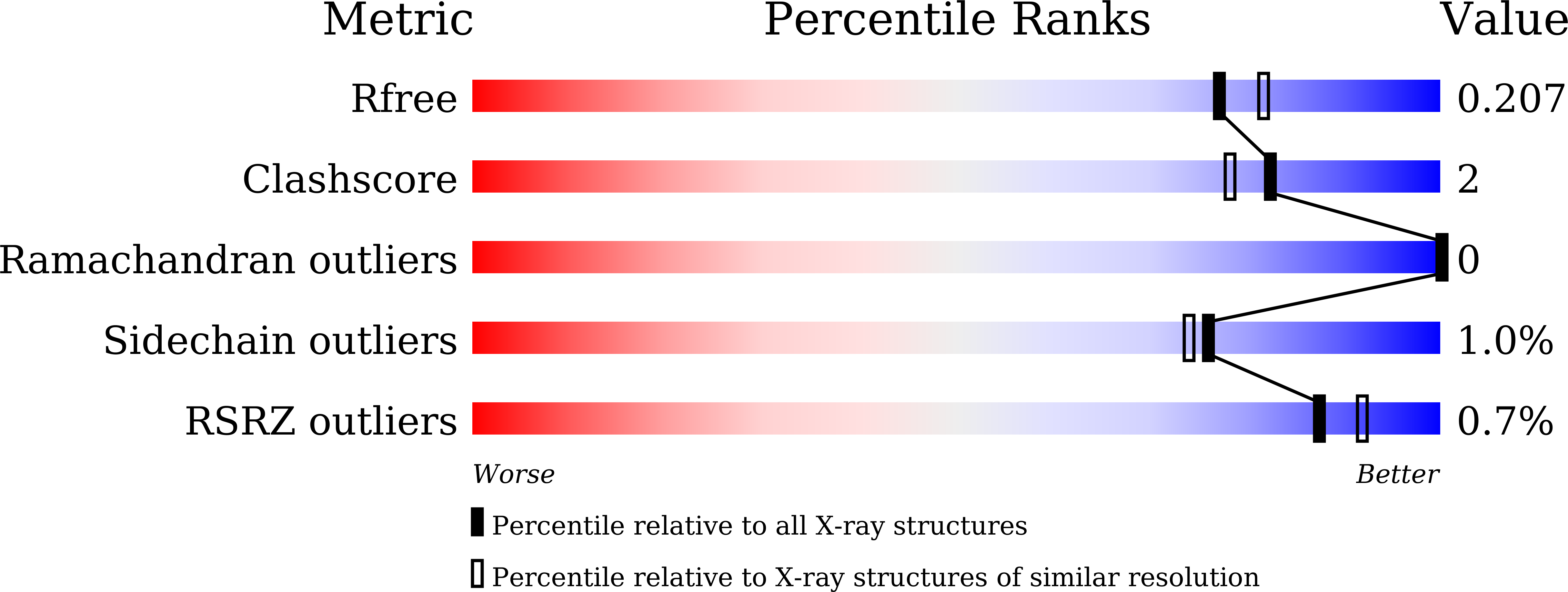
Experimental Data Snapshot
(2021) Adv Synth Catal 363: 4770-4778
Entity ID: 1 | |||||
|---|---|---|---|---|---|
| Molecule | Chains | Sequence Length | Organism | Details | Image |
| D-succinylase | 824 | Cupriavidus sp. P4-10-C | Mutation(s): 1 Gene Names: DSA | 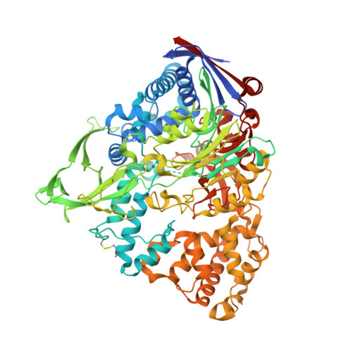 | |
UniProt | |||||
Find proteins for A0A0N7KZ58 (Cupriavidus sp. P4-10-C) Explore A0A0N7KZ58 Go to UniProtKB: A0A0N7KZ58 | |||||
Entity Groups | |||||
| Sequence Clusters | 30% Identity50% Identity70% Identity90% Identity95% Identity100% Identity | ||||
| UniProt Group | A0A0N7KZ58 | ||||
Sequence AnnotationsExpand | |||||
| |||||
| Ligands 6 Unique | |||||
|---|---|---|---|---|---|
| ID | Chains | Name / Formula / InChI Key | 2D Diagram | 3D Interactions | |
| CAD Query on CAD | C [auth A], F [auth A] | CACODYLIC ACID C2 H7 As O2 OGGXGZAMXPVRFZ-UHFFFAOYSA-N |  | ||
| SIN (Subject of Investigation/LOI) Query on SIN | G [auth A], H [auth A], L [auth B], M [auth B], N [auth B] | SUCCINIC ACID C4 H6 O4 KDYFGRWQOYBRFD-UHFFFAOYSA-N |  | ||
| GOL Query on GOL | D [auth A], E [auth A] | GLYCEROL C3 H8 O3 PEDCQBHIVMGVHV-UHFFFAOYSA-N |  | ||
| CO3 Query on CO3 | I [auth A] | CARBONATE ION C O3 BVKZGUZCCUSVTD-UHFFFAOYSA-L |  | ||
| CA Query on CA | J [auth A], O [auth B] | CALCIUM ION Ca BHPQYMZQTOCNFJ-UHFFFAOYSA-N |  | ||
| CL Query on CL | K [auth A], P [auth B] | CHLORIDE ION Cl VEXZGXHMUGYJMC-UHFFFAOYSA-M |  | ||
| Length ( Å ) | Angle ( ˚ ) |
|---|---|
| a = 62.057 | α = 90 |
| b = 129.806 | β = 90 |
| c = 200.68 | γ = 90 |
| Software Name | Purpose |
|---|---|
| PHENIX | refinement |
| DENZO | data reduction |
| SCALA | data scaling |
| PHASER | phasing |
RCSB PDB (citation) is hosted by
RCSB PDB is a member of the

