7E1C

Structure of MreB3 from Spiroplasma eriocheiris


Experimental Data Snapshot

  • Method: X-RAY DIFFRACTION
  • Resolution: 1.90 Å
  • R-Value Free: 0.221 
  • R-Value Work: 0.185 
  • R-Value Observed: 0.188 

wwPDB Validation   3D Report Full Report


This is version 1.2 of the entry. See complete history


Literature

ATP-dependent polymerization dynamics of bacterial actin proteins involved in Spiroplasma swimming.

Takahashi, D.Fujiwara, I.Sasajima, Y.Narita, A.Imada, K.Miyata, M.

(2022) Open Biol 12: 220083-220083

  • DOI: https://doi.org/10.1098/rsob.220083
  • Primary Citation of Related Structures:  
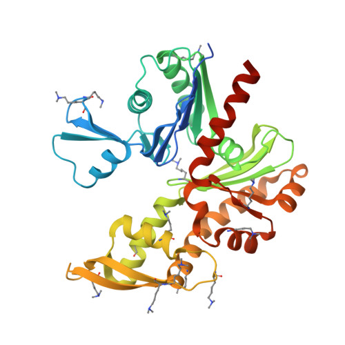
    7E1C, 7E1G

  • PubMed Abstract: 

    MreB is a bacterial protein belonging to the actin superfamily. This protein polymerizes into an antiparallel double-stranded filament that determines cell shape by maintaining cell wall synthesis. Spiroplasma eriocheiris , a helical wall-less bacterium, has five MreB homologous (SpeMreB1-5) that probably contribute to swimming motility. Here, we investigated the structure, ATPase activity and polymerization dynamics of SpeMreB3 and SpeMreB5. SpeMreB3 polymerized into a double-stranded filament with possible antiparallel polarity, while SpeMreB5 formed sheets which contained the antiparallel filament, upon nucleotide binding. SpeMreB3 showed slow P i release owing to the lack of an amino acid motif conserved in the catalytic centre of MreB family proteins. Our SpeMreB3 crystal structures and analyses of SpeMreB3 and SpeMreB5 variants showed that the amino acid motif probably plays a role in eliminating a nucleophilic water proton during ATP hydrolysis. Sedimentation assays suggest that SpeMreB3 has a lower polymerization activity than SpeMreB5, though their polymerization dynamics are qualitatively similar to those of other actin superfamily proteins, in which pre-ATP hydrolysis and post-P i release states are unfavourable for them to remain as filaments.


  • Organizational Affiliation

    Graduate School of Science, Osaka Metropolitan University, Osaka, Japan.


Macromolecules
Find similar proteins by:  (by identity cutoff)  |  3D Structure
Entity ID: 1
MoleculeChains Sequence LengthOrganismDetailsImage
Cell shape-determining protein MreB355Spiroplasma eriocheirisMutation(s): 0 
Gene Names: mreB3mreBSERIO_v1c12260
UniProt
Find proteins for A0A0H3XJK5 (Spiroplasma eriocheiris)
Explore A0A0H3XJK5 
Go to UniProtKB:  A0A0H3XJK5
Entity Groups  
Sequence Clusters30% Identity50% Identity70% Identity90% Identity95% Identity100% Identity
UniProt GroupA0A0H3XJK5
Sequence Annotations
Expand
  • Reference Sequence
Small Molecules
Modified Residues  1 Unique
IDChains TypeFormula2D DiagramParent
MLY
Query on MLY
A
L-PEPTIDE LINKINGC8 H18 N2 O2LYS
Experimental Data & Validation

Experimental Data

  • Method: X-RAY DIFFRACTION
  • Resolution: 1.90 Å
  • R-Value Free: 0.221 
  • R-Value Work: 0.185 
  • R-Value Observed: 0.188 
  • Space Group: P 1 21 1
Unit Cell:
Length ( Å )Angle ( ˚ )
a = 52.42α = 90
b = 68.09β = 91.73
c = 54.57γ = 90
Software Package:
Software NamePurpose
BSSdata collection
PHENIXrefinement
iMOSFLMdata reduction
Aimlessdata scaling
PHASERphasing

Structure Validation

View Full Validation Report


Entry History & Funding Information

Deposition Data


Funding OrganizationLocationGrant Number
Japan Society for the Promotion of Science (JSPS)JapanJP17H01544
Japan Science and TechnologyJapanJPMJCR19S5

Revision History  (Full details and data files)

  • Version 1.0: 2022-02-16
    Type: Initial release
  • Version 1.1: 2023-03-08
    Changes: Database references
  • Version 1.2: 2023-11-29
    Changes: Data collection, Refinement description