7E1B

Crystal structure of VbrR-DNA complex


Experimental Data Snapshot

  • Method: X-RAY DIFFRACTION
  • Resolution: 4.59 Å
  • R-Value Free: 0.321 
  • R-Value Work: 0.265 
  • R-Value Observed: 0.277 

wwPDB Validation   3D Report Full Report


This is version 1.1 of the entry. See complete history


Literature

Structural basis of phosphorylation-induced activation of the response regulator VbrR.

Hong, S.Guo, J.Zhang, X.Zhou, X.Zhang, P.Yu, F.

(2023) Acta Biochim Biophys Sin (Shanghai) 

  • DOI: https://doi.org/10.3724/abbs.2022200
  • Primary Citation of Related Structures:  
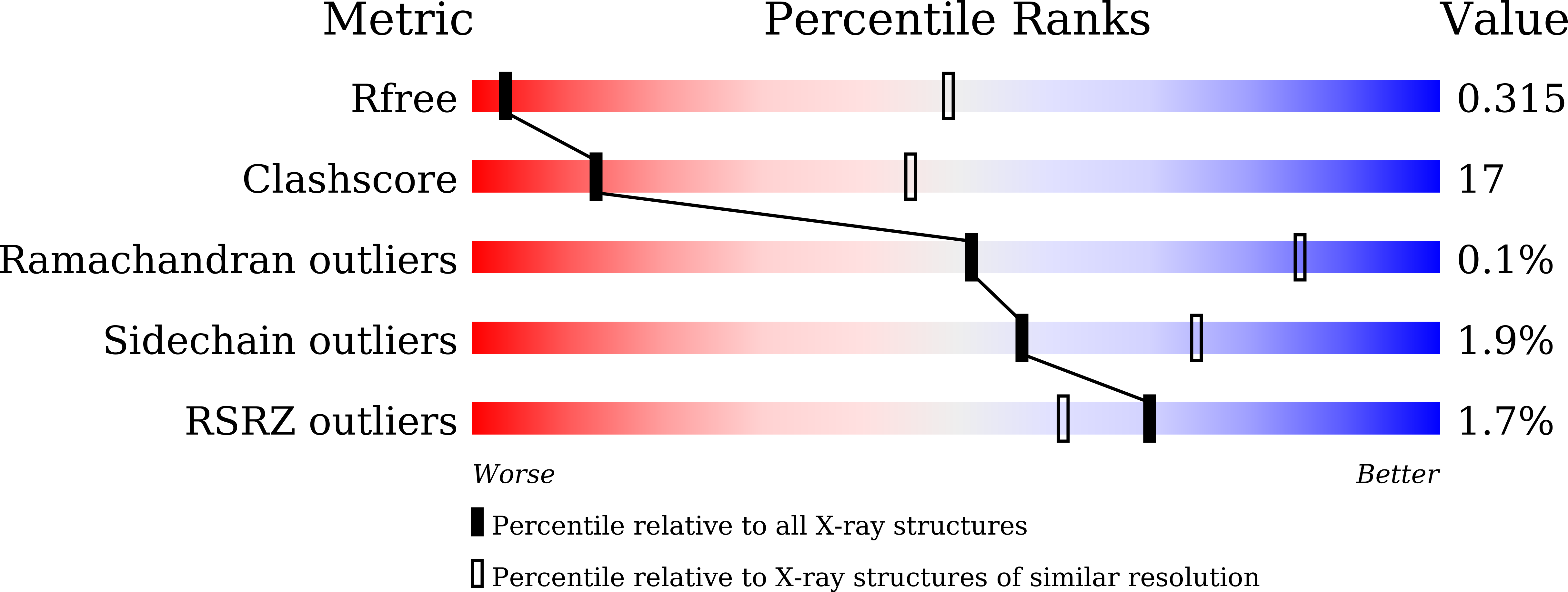
    7E1B, 7E1D, 7E1F, 7E1H

  • PubMed Abstract: 

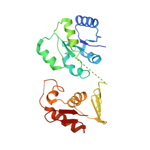
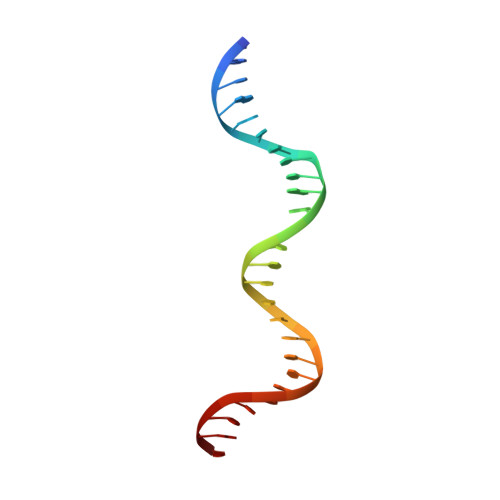
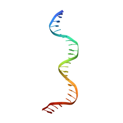
    Two-component systems typically consist of a paired histidine kinase and response regulator and couple environmental changes to adaptive responses. The response regulator VbrR from Vibrio parahaemolyticus , a member of the OmpR/PhoB family, regulates virulence and antibiotic resistance genes. The activation mechanism of VbrR remains unclear. Here, we report the crystal structures of full-length VbrR in complex with DNA in the active conformation and the N-terminal receiver domain (RD) and the C-terminal DNA-binding domain (DBD) in both active and inactive conformations. Structural and biochemical analyses suggest that unphosphorylated VbrR adopts mainly as inactive dimers through the DBD at the autoinhibitory state. The RD undergoes a monomer-to-dimer transition upon phosphorylation, which further induces the transition of DBD from an autoinhibitory dimer to an active dimer and enables its binding with target DNA. Our study suggests a new model for phosphorylation-induced activation of response regulators and sheds light on the pathogenesis of V . parahaemolyticus .


  • Organizational Affiliation

    National Key Laboratory of Plant Molecular Genetics, Center for Excellence in Molecular Plant Sciences, Institute of Plant Physiology and Ecology, Chinese Academy of Sciences, Shanghai 200032, China.


Macromolecules

Find similar proteins by:  (by identity cutoff)  |  3D Structure
Entity ID: 1
MoleculeChains Sequence LengthOrganismDetailsImage
DNA-binding response regulator
A, B, C, D, E
A, B, C, D, E, F, G, H
220Vibrio parahaemolyticusMutation(s): 0 
Gene Names: C9I78_18460CA163_10520CGH73_12770CGI34_20580CGI42_14750D5E78_24805F0L89_03250F0L99_22745WR32_19050
UniProt
Find proteins for Q87HP4 (Vibrio parahaemolyticus serotype O3:K6 (strain RIMD 2210633))
Explore Q87HP4 
Go to UniProtKB:  Q87HP4
Entity Groups  
Sequence Clusters30% Identity50% Identity70% Identity90% Identity95% Identity100% Identity
UniProt GroupQ87HP4
Sequence Annotations
Expand
  • Reference Sequence
Find similar nucleic acids by:  (by identity cutoff)  |  3D Structure
Entity ID: 2
MoleculeChains LengthOrganismImage
DNA (26-MER)I [auth Y],
K [auth I]
26Vibrio parahaemolyticus
Sequence Annotations
Expand
  • Reference Sequence
Find similar nucleic acids by:  (by identity cutoff)  |  3D Structure
Entity ID: 3
MoleculeChains LengthOrganismImage
DNA (26-MER)J [auth Z],
L [auth J]
26Vibrio parahaemolyticus
Sequence Annotations
Expand
  • Reference Sequence
Experimental Data & Validation

Experimental Data

  • Method: X-RAY DIFFRACTION
  • Resolution: 4.59 Å
  • R-Value Free: 0.321 
  • R-Value Work: 0.265 
  • R-Value Observed: 0.277 
  • Space Group: C 1 2 1
Unit Cell:
Length ( Å )Angle ( ˚ )
a = 329.942α = 90
b = 114.064β = 110.02
c = 114.036γ = 90
Software Package:
Software NamePurpose
PHENIXrefinement
PDB_EXTRACTdata extraction
HKL-3000data reduction
HKL-3000data scaling
PHENIXphasing

Structure Validation

View Full Validation Report


Entry History & Funding Information

Deposition Data


Funding OrganizationLocationGrant Number
National Natural Science Foundation of China (NSFC)China32025020, 31971120

Revision History  (Full details and data files)

  • Version 1.0: 2022-02-09
    Type: Initial release
  • Version 1.1: 2023-02-15
    Changes: Database references