


Crystal structure of the phage-encoded N-acetyltransferase in complex with acetyl-CoA
Hyun, Y., Oh, H.-M., Ha, N.-C.To be published.
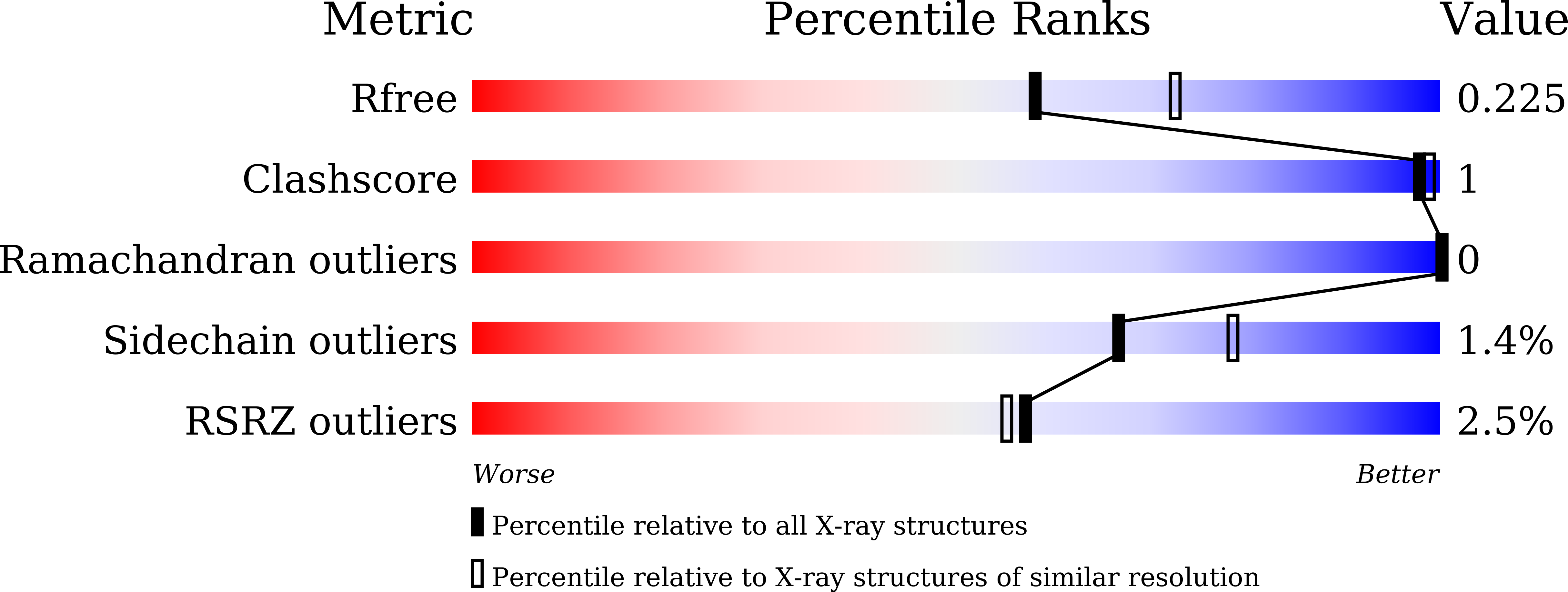
Experimental Data Snapshot
Entity ID: 1 | |||||
|---|---|---|---|---|---|
| Molecule | Chains | Sequence Length | Organism | Details | Image |
| Putative acetyltransferase | 186 | Salmonella phage SPN3US | Mutation(s): 0 Gene Names: SPN3US_0088 | 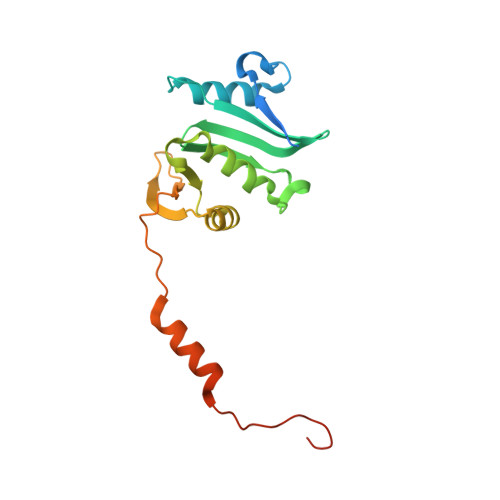 | |
UniProt | |||||
Find proteins for G5DEI1 (Salmonella phage SPN3US) Explore G5DEI1 Go to UniProtKB: G5DEI1 | |||||
Entity Groups | |||||
| Sequence Clusters | 30% Identity50% Identity70% Identity90% Identity95% Identity100% Identity | ||||
| UniProt Group | G5DEI1 | ||||
Sequence AnnotationsExpand | |||||
| |||||
| Ligands 1 Unique | |||||
|---|---|---|---|---|---|
| ID | Chains | Name / Formula / InChI Key | 2D Diagram | 3D Interactions | |
| ACO (Subject of Investigation/LOI) Query on ACO | B [auth A] | ACETYL COENZYME *A C23 H38 N7 O17 P3 S ZSLZBFCDCINBPY-ZSJPKINUSA-N |  | ||
| Length ( Å ) | Angle ( ˚ ) |
|---|---|
| a = 68.458 | α = 90 |
| b = 68.458 | β = 90 |
| c = 218.964 | γ = 120 |
| Software Name | Purpose |
|---|---|
| PHENIX | refinement |
| HKL-2000 | data reduction |
| HKL-2000 | data scaling |
| PHENIX | model building |
| PHENIX | phasing |
RCSB PDB (citation) is hosted by
RCSB PDB is a member of the

