7DDK

Crystal structures of Na+,K+-ATPase in complex with rostafuroxin


Experimental Data Snapshot

  • Method: X-RAY DIFFRACTION
  • Resolution: 3.50 Å
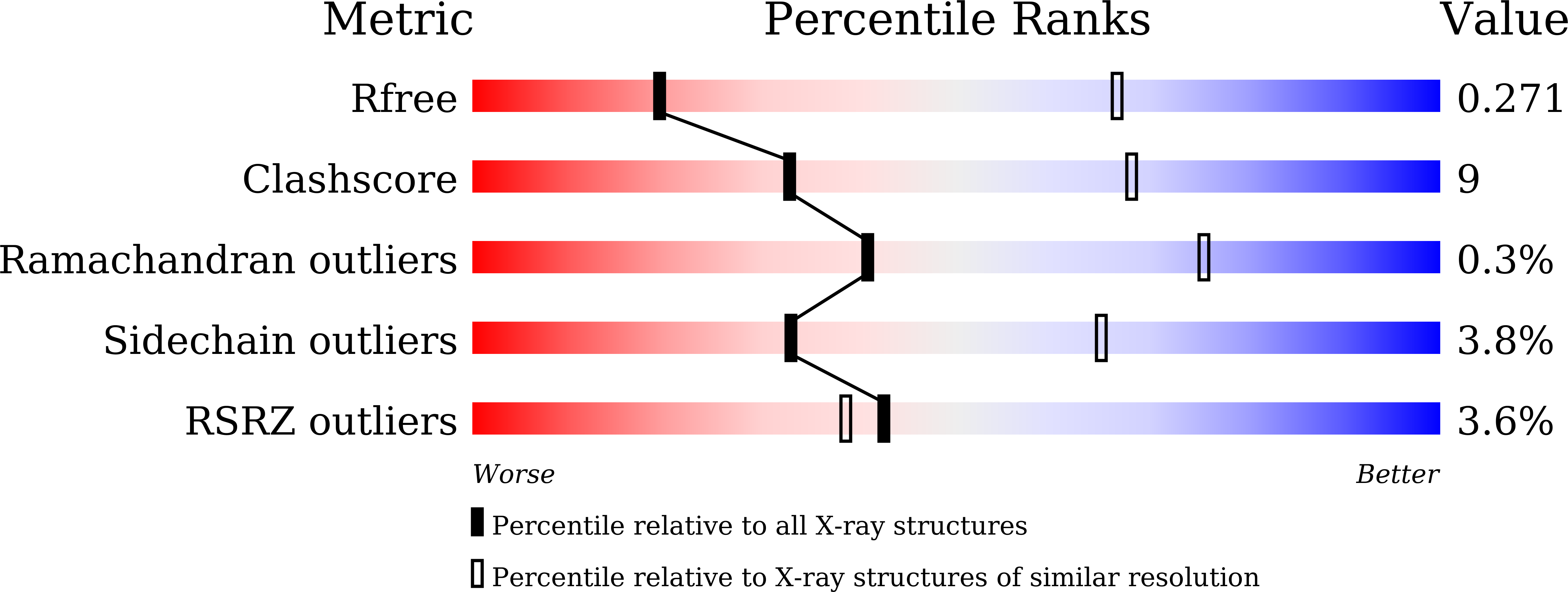
  • R-Value Free: 0.269 
  • R-Value Work: 0.220 
  • R-Value Observed: 0.222 

Starting Model: experimental
View more details

wwPDB Validation   3D Report Full Report


Ligand Structure Quality Assessment 


This is version 1.1 of the entry. See complete history

Re-refinement Note

This entry reflects an alternative modeling of the original data in: 6KQ0


Literature

Binding of cardiotonic steroids to Na + ,K + -ATPase in the E2P state.

Kanai, R.Cornelius, F.Ogawa, H.Motoyama, K.Vilsen, B.Toyoshima, C.

(2021) Proc Natl Acad Sci U S A 118

  • DOI: https://doi.org/10.1073/pnas.2020438118
  • Primary Citation of Related Structures:  
    7D91, 7D92, 7D93, 7D94, 7DDF, 7DDH, 7DDI, 7DDK, 7DDL

  • PubMed Abstract: 

    The sodium pump (Na + , K + -ATPase, NKA) is vital for animal cells, as it actively maintains Na + and K + electrochemical gradients across the cell membrane. It is a target of cardiotonic steroids (CTSs) such as ouabain and digoxin. As CTSs are almost unique strong inhibitors specific to NKA, a wide range of derivatives has been developed for potential therapeutic use. Several crystal structures have been published for NKA-CTS complexes, but they fail to explain the largely different inhibitory properties of the various CTSs. For instance, although CTSs are thought to inhibit ATPase activity by binding to NKA in the E2P state, we do not know if large conformational changes accompany binding, as no crystal structure is available for the E2P state free of CTS. Here, we describe crystal structures of the BeF 3 - complex of NKA representing the E2P ground state and then eight crystal structures of seven CTSs, including rostafuroxin and istaroxime, two new members under clinical trials, in complex with NKA in the E2P state. The conformations of NKA are virtually identical in all complexes with and without CTSs, showing that CTSs bind to a preformed cavity in NKA. By comparing the inhibitory potency of the CTSs measured under four different conditions, we elucidate how different structural features of the CTSs result in different inhibitory properties. The crystal structures also explain K + -antagonism and suggest a route to isoform specific CTSs.


  • Organizational Affiliation

    Institute for Quantitative Biosciences, The University of Tokyo, 113-0032 Tokyo, Japan.


Macromolecules
Find similar proteins by:  (by identity cutoff)  |  3D Structure
Entity ID: 1
MoleculeChains Sequence LengthOrganismDetailsImage
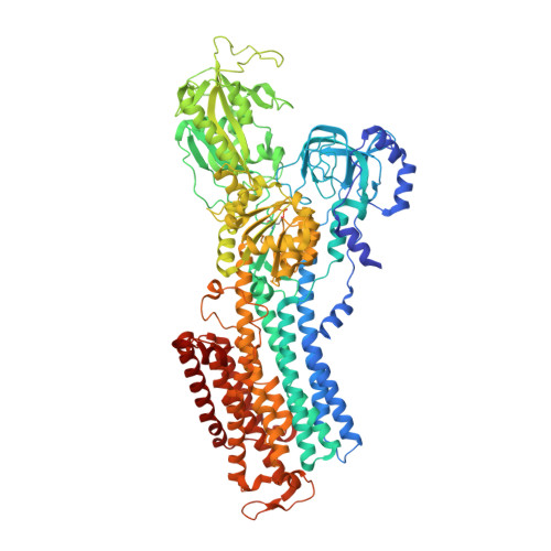
Sodium/potassium-transporting ATPase subunit alpha-1A,
D [auth C]
1,016Sus scrofaMutation(s): 0 
EC: 7.2.2.13
Membrane Entity: Yes 
UniProt
Find proteins for P05024 (Sus scrofa)
Explore P05024 
Go to UniProtKB:  P05024
Entity Groups  
Sequence Clusters30% Identity50% Identity70% Identity90% Identity95% Identity100% Identity
UniProt GroupP05024
Sequence Annotations
Expand
  • Reference Sequence
Find similar proteins by:  (by identity cutoff)  |  3D Structure
Entity ID: 2
MoleculeChains Sequence LengthOrganismDetailsImage
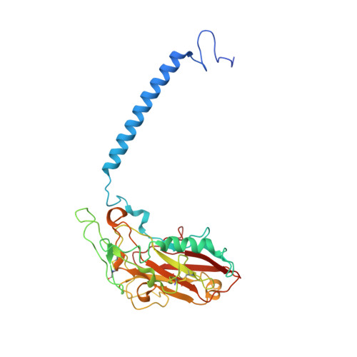
Sodium/potassium-transporting ATPase subunit beta-1B,
E [auth D]
303Sus scrofaMutation(s): 0 
Membrane Entity: Yes 
UniProt
Find proteins for P05027 (Sus scrofa)
Explore P05027 
Go to UniProtKB:  P05027
Entity Groups  
Sequence Clusters30% Identity50% Identity70% Identity90% Identity95% Identity100% Identity
UniProt GroupP05027
Glycosylation
Glycosylation Sites: 3
Sequence Annotations
Expand
  • Reference Sequence
Find similar proteins by:  (by identity cutoff)  |  3D Structure
Entity ID: 3
MoleculeChains Sequence LengthOrganismDetailsImage
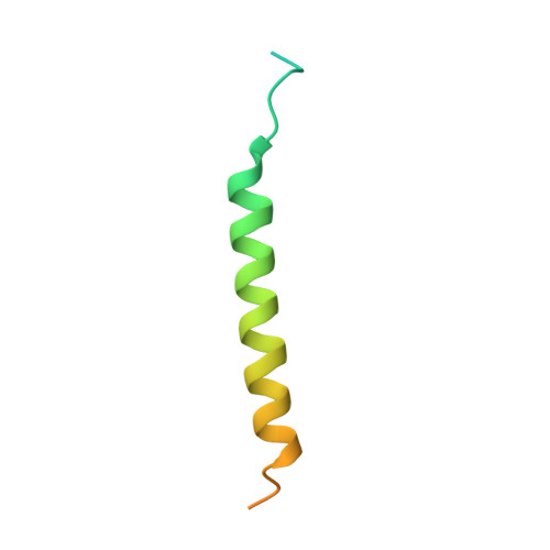
FXYD domain-containing ion transport regulatorC [auth G],
F [auth E]
65Sus scrofaMutation(s): 0 
Membrane Entity: Yes 
UniProt
Find proteins for Q58K79 (Sus scrofa)
Explore Q58K79 
Go to UniProtKB:  Q58K79
Entity Groups  
Sequence Clusters30% Identity50% Identity70% Identity90% Identity95% Identity100% Identity
UniProt GroupQ58K79
Sequence Annotations
Expand
  • Reference Sequence
Oligosaccharides

Help

Entity ID: 4
MoleculeChains Length2D Diagram Glycosylation3D Interactions
2-acetamido-2-deoxy-beta-D-glucopyranose-(1-4)-2-acetamido-2-deoxy-beta-D-glucopyranoseG [auth F],
H,
I,
J
2N-Glycosylation
Glycosylation Resources
GlyTouCan:  G42666HT
GlyCosmos:  G42666HT
GlyGen:  G42666HT
Small Molecules
Ligands 6 Unique
IDChains Name / Formula / InChI Key2D Diagram3D Interactions
PCW
Query on PCW

Download Ideal Coordinates CCD File 
CA [auth C]
DA [auth C]
EA [auth C]
FA [auth C]
GA [auth C]
CA [auth C],
DA [auth C],
EA [auth C],
FA [auth C],
GA [auth C],
HA [auth C],
IA [auth C],
N [auth A],
O [auth A],
P [auth A],
Q [auth A],
R [auth A],
S [auth A]
1,2-DIOLEOYL-SN-GLYCERO-3-PHOSPHOCHOLINE
C44 H85 N O8 P
SNKAWJBJQDLSFF-NVKMUCNASA-O
CLR
Query on CLR

Download Ideal Coordinates CCD File 
BA [auth C]
LA [auth D]
MA [auth E]
U [auth A]
W [auth B]
BA [auth C],
LA [auth D],
MA [auth E],
U [auth A],
W [auth B],
X [auth G]
CHOLESTEROL
C27 H46 O
HVYWMOMLDIMFJA-DPAQBDIFSA-N
E4R (Subject of Investigation/LOI)
Query on E4R

Download Ideal Coordinates CCD File 
JA [auth C],
T [auth A]
(3S,5R,8R,9S,10S,13S,14S,17S)-17-(furan-3-yl)-10,13-dimethyl-2,3,4,5,6,7,8,9,11,12,15,16-dodecahydro-1H-cyclopenta[a]phenanthrene-3,14,17-triol
C23 H34 O4
AEAPORIZZWBIEX-DTBDINHYSA-N
NAG
Query on NAG

Download Ideal Coordinates CCD File 
KA [auth D],
V [auth B]
2-acetamido-2-deoxy-beta-D-glucopyranose
C8 H15 N O6
OVRNDRQMDRJTHS-FMDGEEDCSA-N
MG
Query on MG

Download Ideal Coordinates CCD File 
AA [auth C],
K [auth A],
M [auth A],
Y [auth C]
MAGNESIUM ION
Mg
JLVVSXFLKOJNIY-UHFFFAOYSA-N
NA
Query on NA

Download Ideal Coordinates CCD File 
L [auth A],
Z [auth C]
SODIUM ION
Na
FKNQFGJONOIPTF-UHFFFAOYSA-N
Modified Residues  1 Unique
IDChains TypeFormula2D DiagramParent
PHD
Query on PHD
A,
D [auth C]
L-PEPTIDE LINKINGC4 H8 N O7 PASP
Experimental Data & Validation

Experimental Data

  • Method: X-RAY DIFFRACTION
  • Resolution: 3.50 Å
  • R-Value Free: 0.269 
  • R-Value Work: 0.220 
  • R-Value Observed: 0.222 
  • Space Group: P 21 21 21
Unit Cell:
Length ( Å )Angle ( ˚ )
a = 114.357α = 90
b = 118.176β = 90
c = 494.668γ = 90
Software Package:
Software NamePurpose
HKL-2000data reduction
SCALEPACKdata scaling
MOLREPphasing
PHENIXrefinement

Structure Validation

View Full Validation Report


Ligand Structure Quality Assessment 


Entry History & Funding Information

Deposition Data


Funding OrganizationLocationGrant Number
Japan Society for the Promotion of Science (JSPS)Japan16K18500
Japan Society for the Promotion of Science (JSPS)Japan16H02499
Japan Society for the Promotion of Science (JSPS)Japan19H00975

Revision History  (Full details and data files)

  • Version 1.0: 2021-01-27
    Type: Initial release
  • Version 1.1: 2023-11-29
    Changes: Data collection, Database references, Refinement description