7AQ1

Crystal structure of human mature meprin beta in complex with the specific inhibitor MWT-S-270


Experimental Data Snapshot

  • Method: X-RAY DIFFRACTION
  • Resolution: 2.41 Å
  • R-Value Free: 0.236 
  • R-Value Work: 0.199 
  • R-Value Observed: 0.200 

wwPDB Validation   3D Report Full Report


Ligand Structure Quality Assessment 


This is version 1.2 of the entry. See complete history


Literature

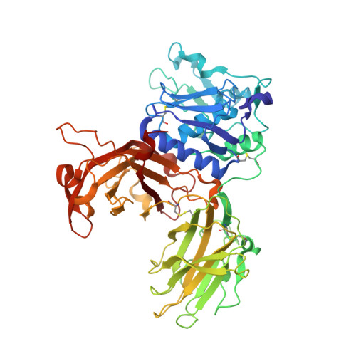
Structure and Dynamics of Meprin beta in Complex with a Hydroxamate-Based Inhibitor.

Linnert, M.Fritz, C.Jager, C.Schlenzig, D.Ramsbeck, D.Kleinschmidt, M.Wermann, M.Demuth, H.U.Parthier, C.Schilling, S.

(2021) Int J Mol Sci 22

  • DOI: https://doi.org/10.3390/ijms22115651
  • Primary Citation of Related Structures:  
    7AQ1

  • PubMed Abstract: 

    The astacin protease Meprin β represents an emerging target for drug development due to its potential involvement in disorders such as acute and chronic kidney injury and fibrosis. Here, we elaborate on the structural basis of inhibition by a specific Meprin β inhibitor. Our analysis of the crystal structure suggests different binding modes of the inhibitor to the active site. This flexibility is caused, at least in part, by movement of the C-terminal region of the protease domain (CTD). The CTD movement narrows the active site cleft upon inhibitor binding. Compared with other astacin proteases, among these the highly homologous isoenzyme Meprin α, differences in the subsites account for the unique selectivity of the inhibitor. Although the inhibitor shows substantial flexibility in orientation within the active site, the structural data as well as binding analyses, including molecular dynamics simulations, support a contribution of electrostatic interactions, presumably by arginine residues, to binding and specificity. Collectively, the results presented here and previously support an induced fit and substantial movement of the CTD upon ligand binding and, possibly, during catalysis. To the best of our knowledge, we here present the first structure of a Meprin β holoenzyme containing a zinc ion and a specific inhibitor bound to the active site. The structural data will guide rational drug design and the discovery of highly potent Meprin inhibitors.


  • Organizational Affiliation

    Fraunhofer Institute for Cell Therapy and Immunology, Department of Drug Design and Target Validation, Weinbergweg 22, 06120 Halle (Saale), Germany.


Macromolecules
Find similar proteins by:  (by identity cutoff)  |  3D Structure
Entity ID: 1
MoleculeChains Sequence LengthOrganismDetailsImage
Meprin B subunit beta
A, B
534Homo sapiensMutation(s): 0 
Gene Names: MEP1B
EC: 3.4.24.63
UniProt & NIH Common Fund Data Resources
Find proteins for Q16820 (Homo sapiens)
Explore Q16820 
Go to UniProtKB:  Q16820
PHAROS:  Q16820
GTEx:  ENSG00000141434 
Entity Groups  
Sequence Clusters30% Identity50% Identity70% Identity90% Identity95% Identity100% Identity
UniProt GroupQ16820
Sequence Annotations
Expand
  • Reference Sequence
Oligosaccharides

Help

Entity ID: 2
MoleculeChains Length2D Diagram Glycosylation3D Interactions
2-acetamido-2-deoxy-beta-D-glucopyranose-(1-4)-2-acetamido-2-deoxy-beta-D-glucopyranoseC [auth H],
E [auth K]
2N-Glycosylation
Glycosylation Resources
GlyTouCan:  G42666HT
GlyCosmos:  G42666HT
GlyGen:  G42666HT
Entity ID: 3
MoleculeChains Length2D Diagram Glycosylation3D Interactions
alpha-D-mannopyranose-(1-2)-alpha-D-mannopyranose-(1-6)-[alpha-D-mannopyranose-(1-3)]alpha-D-mannopyranose-(1-6)-[alpha-D-mannopyranose-(1-2)-alpha-D-mannopyranose-(1-3)]beta-D-mannopyranose-(1-4)-2-acetamido-2-deoxy-beta-D-glucopyranose-(1-4)-2-acetamido-2-deoxy-beta-D-glucopyranoseD [auth I]9N-Glycosylation
Glycosylation Resources
GlyTouCan:  G83161QT
GlyCosmos:  G83161QT
GlyGen:  G83161QT
Entity ID: 4
MoleculeChains Length2D Diagram Glycosylation3D Interactions
alpha-D-mannopyranose-(1-2)-alpha-D-mannopyranose-(1-6)-[alpha-D-mannopyranose-(1-3)]alpha-D-mannopyranose-(1-6)-[alpha-D-mannopyranose-(1-3)]beta-D-mannopyranose-(1-4)-2-acetamido-2-deoxy-beta-D-glucopyranose-(1-4)-2-acetamido-2-deoxy-beta-D-glucopyranoseF [auth L]8N-Glycosylation
Glycosylation Resources
GlyTouCan:  G61846BY
GlyCosmos:  G61846BY
GlyGen:  G61846BY
Small Molecules
Ligands 6 Unique
IDChains Name / Formula / InChI Key2D Diagram3D Interactions
RUE (Subject of Investigation/LOI)
Query on RUE

Download Ideal Coordinates CCD File 
K [auth A],
U [auth B]
3-[[(3-Carboxyphenyl)methyl-[2-(hydroxyamino)-2-oxoethyl]amino]methyl]benzoic acid
C18 H18 N2 O6
KHSBLMDTVROJFV-UHFFFAOYSA-N
NAG
Query on NAG

Download Ideal Coordinates CCD File 
O [auth A],
P [auth A],
Q [auth A],
Y [auth B],
Z [auth B]
2-acetamido-2-deoxy-beta-D-glucopyranose
C8 H15 N O6
OVRNDRQMDRJTHS-FMDGEEDCSA-N
ZN
Query on ZN

Download Ideal Coordinates CCD File 
G [auth A],
R [auth B]
ZINC ION
Zn
PTFCDOFLOPIGGS-UHFFFAOYSA-N
EDO
Query on EDO

Download Ideal Coordinates CCD File 
H [auth A],
I [auth A],
J [auth A],
S [auth B],
T [auth B]
1,2-ETHANEDIOL
C2 H6 O2
LYCAIKOWRPUZTN-UHFFFAOYSA-N
CA
Query on CA

Download Ideal Coordinates CCD File 
L [auth A],
M [auth A],
V [auth B],
W [auth B]
CALCIUM ION
Ca
BHPQYMZQTOCNFJ-UHFFFAOYSA-N
CL
Query on CL

Download Ideal Coordinates CCD File 
N [auth A],
X [auth B]
CHLORIDE ION
Cl
VEXZGXHMUGYJMC-UHFFFAOYSA-M
Experimental Data & Validation

Experimental Data

  • Method: X-RAY DIFFRACTION
  • Resolution: 2.41 Å
  • R-Value Free: 0.236 
  • R-Value Work: 0.199 
  • R-Value Observed: 0.200 
  • Space Group: C 1 2 1
Unit Cell:
Length ( Å )Angle ( ˚ )
a = 162.25α = 90
b = 72.44β = 118.43
c = 135.47γ = 90
Software Package:
Software NamePurpose
PHENIXrefinement
XDSdata reduction
XSCALEdata scaling
PHASERphasing
PDB_EXTRACTdata extraction

Structure Validation

View Full Validation Report


Ligand Structure Quality Assessment 


Entry History 

Deposition Data

Revision History  (Full details and data files)

  • Version 1.0: 2021-03-03
    Type: Initial release
  • Version 1.1: 2021-06-16
    Changes: Database references
  • Version 1.2: 2024-01-31
    Changes: Data collection, Database references, Refinement description