7ACY

H/L (SLPH/SLPL) complex from C. difficile (CD630 strain)


Experimental Data Snapshot

  • Method: X-RAY DIFFRACTION
  • Resolution: 2.55 Å
  • R-Value Free: 0.260 
  • R-Value Work: 0.230 
  • R-Value Observed: 0.230 

wwPDB Validation   3D Report Full Report


This is version 1.1 of the entry. See complete history


Literature

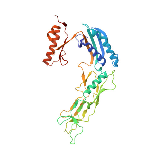
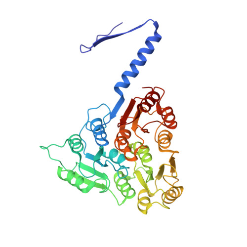
Structure and assembly of the S-layer in C. difficile.

Lanzoni-Mangutchi, P.Banerji, O.Wilson, J.Barwinska-Sendra, A.Kirk, J.A.Vaz, F.O'Beirne, S.Basle, A.El Omari, K.Wagner, A.Fairweather, N.F.Douce, G.R.Bullough, P.A.Fagan, R.P.Salgado, P.S.

(2022) Nat Commun 13: 970-970

  • DOI: https://doi.org/10.1038/s41467-022-28196-w
  • Primary Citation of Related Structures:  
    7ACV, 7ACW, 7ACX, 7ACY, 7ACZ, 7QGQ

  • PubMed Abstract: 

    Many bacteria and archaea possess a two-dimensional protein array, or S-layer, that covers the cell surface and plays crucial roles in cell physiology. Here, we report the crystal structure of SlpA, the main S-layer protein of the bacterial pathogen Clostridioides difficile, and use electron microscopy to study S-layer organisation and assembly. The SlpA crystal lattice mimics S-layer assembly in the cell, through tiling of triangular prisms above the cell wall, interlocked by distinct ridges facing the environment. Strikingly, the array is very compact, with pores of only ~10 Å in diameter, compared to other S-layers (30-100 Å). The surface-exposed flexible ridges are partially dispensable for overall structure and assembly, although a mutant lacking this region becomes susceptible to lysozyme, an important molecule in host defence. Thus, our work gives insights into S-layer organisation and provides a basis for development of C. difficile-specific therapeutics.


  • Organizational Affiliation

    Biosciences Institute, Faculty of Medical Sciences, Newcastle University, Newcastle upon Tyne, UK.


Macromolecules
Find similar proteins by:  (by identity cutoff)  |  3D Structure
Entity ID: 1
MoleculeChains Sequence LengthOrganismDetailsImage
S-layer protein
A, C
318Clostridioides difficile 630Mutation(s): 0 
UniProt
Find proteins for Q183M8 (Clostridioides difficile (strain 630))
Explore Q183M8 
Go to UniProtKB:  Q183M8
Entity Groups  
Sequence Clusters30% Identity50% Identity70% Identity90% Identity95% Identity100% Identity
UniProt GroupQ183M8
Sequence Annotations
Expand
  • Reference Sequence
Find similar proteins by:  (by identity cutoff)  |  3D Structure
Entity ID: 2
MoleculeChains Sequence LengthOrganismDetailsImage
S-layer protein
B, D
373Clostridioides difficile 630Mutation(s): 0 
UniProt
Find proteins for Q183M8 (Clostridioides difficile (strain 630))
Explore Q183M8 
Go to UniProtKB:  Q183M8
Entity Groups  
Sequence Clusters30% Identity50% Identity70% Identity90% Identity95% Identity100% Identity
UniProt GroupQ183M8
Sequence Annotations
Expand
  • Reference Sequence
Experimental Data & Validation

Experimental Data

  • Method: X-RAY DIFFRACTION
  • Resolution: 2.55 Å
  • R-Value Free: 0.260 
  • R-Value Work: 0.230 
  • R-Value Observed: 0.230 
  • Space Group: P 1
Unit Cell:
Length ( Å )Angle ( ˚ )
a = 72.67α = 81.89
b = 78.25β = 66.98
c = 81.58γ = 65.26
Software Package:
Software NamePurpose
REFMACrefinement
DIALSdata reduction
Aimlessdata scaling
PHASERphasing

Structure Validation

View Full Validation Report


Entry History & Funding Information

Deposition Data


Funding OrganizationLocationGrant Number
Wellcome TrustUnited Kingdom204877/Z/16/Z

Revision History  (Full details and data files)

  • Version 1.0: 2022-03-09
    Type: Initial release
  • Version 1.1: 2024-01-31
    Changes: Data collection, Refinement description