


ZK177.8 of Caenorhabditis elegans: Aicardi-Goutieres Syndrome SAMHD1 Ortholog
Maehigashi, T., Lim, C., Wade, L.R., Bowen, N., Knecht, K., Schinazi, R.F., Xiong, Y., Kim, B.To be published.
Experimental Data Snapshot
Entity ID: 1 | |||||
|---|---|---|---|---|---|
| Molecule | Chains | Sequence Length | Organism | Details | Image |
| ZK177.8 | A, B, C [auth D], D [auth C] | 526 | Caenorhabditis elegans | Mutation(s): 2 Gene Names: ZK177.8 | 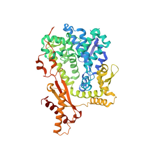 |
UniProt | |||||
Find proteins for Q09374 (Caenorhabditis elegans) Explore Q09374 Go to UniProtKB: Q09374 | |||||
Entity Groups | |||||
| Sequence Clusters | 30% Identity50% Identity70% Identity90% Identity95% Identity100% Identity | ||||
| UniProt Group | Q09374 | ||||
Sequence AnnotationsExpand | |||||
| |||||
| Ligands 4 Unique | |||||
|---|---|---|---|---|---|
| ID | Chains | Name / Formula / InChI Key | 2D Diagram | 3D Interactions | |
| GTP (Subject of Investigation/LOI) Query on GTP | F [auth A], M [auth B], T [auth D], W [auth C] | GUANOSINE-5'-TRIPHOSPHATE C10 H16 N5 O14 P3 XKMLYUALXHKNFT-UUOKFMHZSA-N |  | ||
| DTP (Subject of Investigation/LOI) Query on DTP | E [auth A] G [auth A] L [auth B] N [auth B] S [auth D] | 2'-DEOXYADENOSINE 5'-TRIPHOSPHATE C10 H16 N5 O12 P3 SUYVUBYJARFZHO-RRKCRQDMSA-N |  | ||
| SIN Query on SIN | J [auth A] K [auth A] P [auth B] Q [auth B] Y [auth C] | SUCCINIC ACID C4 H6 O4 KDYFGRWQOYBRFD-UHFFFAOYSA-N |  | ||
| MG Query on MG | H [auth A], I [auth A], O [auth B], R [auth D] | MAGNESIUM ION Mg JLVVSXFLKOJNIY-UHFFFAOYSA-N |  | ||
| Length ( Å ) | Angle ( ˚ ) |
|---|---|
| a = 90.681 | α = 65.58 |
| b = 90.557 | β = 65.49 |
| c = 93.357 | γ = 86.22 |
| Software Name | Purpose |
|---|---|
| SCALEPACK | data scaling |
| REFMAC | refinement |
| PDB_EXTRACT | data extraction |
| HKL-2000 | data reduction |
| BUCCANEER | phasing |
| Funding Organization | Location | Grant Number |
|---|---|---|
| National Institutes of Health/National Institute of General Medical Sciences (NIH/NIGMS) | United States | AI150451 |
| National Institutes of Health/National Human Genome Research Institute (NIH/NHGRI) | United States | AI136581 |
RCSB PDB (citation) is hosted by
RCSB PDB is a member of the

