


Crystal Structure of Enolase from Chlamydia trachomatis
Dranow, D.M., Mayclin, S.J., Lorimer, D.D., Horanyi, P.S., Edwards, T.E.To be published.
Experimental Data Snapshot
wwPDB Validation 3D Report Full Report

Entity ID: 1 | |||||
|---|---|---|---|---|---|
| Molecule | Chains | Sequence Length | Organism | Details | Image |
| Enolase | 432 | Chlamydia trachomatis L2b/UCH-1/proctitis | Mutation(s): 0 Gene Names: eno, CTLon_0844 EC: 4.2.1.11 | 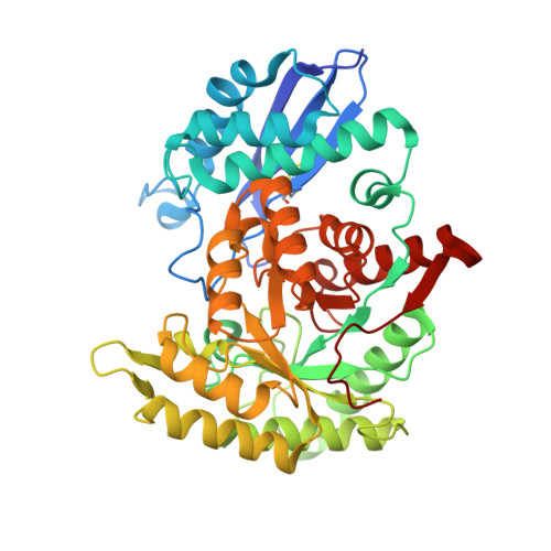 | |
UniProt | |||||
Find proteins for B0BA40 (Chlamydia trachomatis serovar L2b (strain UCH-1/proctitis)) Explore B0BA40 Go to UniProtKB: B0BA40 | |||||
Entity Groups | |||||
| Sequence Clusters | 30% Identity50% Identity70% Identity90% Identity95% Identity100% Identity | ||||
| UniProt Group | B0BA40 | ||||
Sequence AnnotationsExpand | |||||
| |||||
| Ligands 5 Unique | |||||
|---|---|---|---|---|---|
| ID | Chains | Name / Formula / InChI Key | 2D Diagram | 3D Interactions | |
| MRD Query on MRD | C [auth A], I [auth A], Q [auth B], U [auth B] | (4R)-2-METHYLPENTANE-2,4-DIOL C6 H14 O2 SVTBMSDMJJWYQN-RXMQYKEDSA-N |  | ||
| PO4 Query on PO4 | F [auth A], R [auth B] | PHOSPHATE ION O4 P NBIIXXVUZAFLBC-UHFFFAOYSA-K |  | ||
| EDO Query on EDO | D [auth A] E [auth A] J [auth A] K [auth A] L [auth A] | 1,2-ETHANEDIOL C2 H6 O2 LYCAIKOWRPUZTN-UHFFFAOYSA-N |  | ||
| CL Query on CL | N [auth A] | CHLORIDE ION Cl VEXZGXHMUGYJMC-UHFFFAOYSA-M |  | ||
| MG Query on MG | G [auth A], H [auth A], M [auth A], S [auth B], T [auth B] | MAGNESIUM ION Mg JLVVSXFLKOJNIY-UHFFFAOYSA-N |  | ||
| Length ( Å ) | Angle ( ˚ ) |
|---|---|
| a = 164.28 | α = 90 |
| b = 164.28 | β = 90 |
| c = 89.79 | γ = 90 |
| Software Name | Purpose |
|---|---|
| XDS | data reduction |
| XSCALE | data scaling |
| PHENIX | refinement |
| PDB_EXTRACT | data extraction |
| MoRDa | phasing |
RCSB PDB (citation) is hosted by
RCSB PDB is a member of the

