6HR7

HMG-CoA reductase from Methanothermococcus thermolithotrophicus apo form at 2.4 A resolution


Experimental Data Snapshot

  • Method: X-RAY DIFFRACTION
  • Resolution: 2.40 Å
  • R-Value Free: 0.220 
  • R-Value Work: 0.173 
  • R-Value Observed: 0.175 

wwPDB Validation   3D Report Full Report


This is version 1.2 of the entry. See complete history


Literature

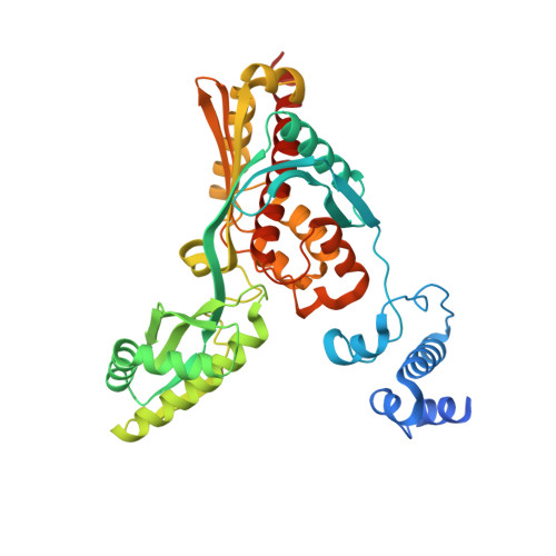
Crystal structure of archaeal HMG-CoA reductase: insights into structural changes of the C-terminal helix of the class-I enzyme.

Vogeli, B.Shima, S.Erb, T.J.Wagner, T.

(2019) FEBS Lett 593: 543-553

  • DOI: https://doi.org/10.1002/1873-3468.13331
  • Primary Citation of Related Structures:  
    6HR7, 6HR8

  • PubMed Abstract: 

    3-hydroxy-3-methylglutaryl-CoA reductase (HMGR) catalyses the last step in mevalonate biosynthesis. HMGR is the target of statin inhibitors that regulate cholesterol concentration in human blood. Here, we report the properties and structures of HMGR from an archaeon Methanothermococcus thermolithotrophicus (mHMGR). The structures of the apoenzyme and the NADPH complex are highly similar to those of human HMGR. A notable exception is C-terminal helix (Lα10-11) that is straight in both mHMGR structures. This helix is kinked and closes the active site in the human enzyme ternary complex, pointing to a substrate-induced structural rearrangement of C-terminal in class-I HMGRs during the catalytic cycle.


  • Organizational Affiliation

    Department of Biochemistry and Synthetic Metabolism, Max Planck Institute for Terrestrial Microbiology, Marburg, Germany.


Macromolecules
Find similar proteins by:  (by identity cutoff)  |  3D Structure
Entity ID: 1
MoleculeChains Sequence LengthOrganismDetailsImage
HMG-CoA reductase
A, B
427Methanothermococcus thermolithotrophicus DSM 2095Mutation(s): 0 
Gene Names: HMGR
EC: 1.1.1.34
UniProt
Find proteins for A0A4V8GZY0 (Methanothermococcus thermolithotrophicus DSM 2095)
Explore A0A4V8GZY0 
Go to UniProtKB:  A0A4V8GZY0
Entity Groups  
Sequence Clusters30% Identity50% Identity70% Identity90% Identity95% Identity100% Identity
UniProt GroupA0A4V8GZY0
Sequence Annotations
Expand
  • Reference Sequence
Small Molecules
Ligands 6 Unique
IDChains Name / Formula / InChI Key2D Diagram3D Interactions
P6G
Query on P6G

Download Ideal Coordinates CCD File 
K [auth A]HEXAETHYLENE GLYCOL
C12 H26 O7
IIRDTKBZINWQAW-UHFFFAOYSA-N
DTT
Query on DTT

Download Ideal Coordinates CCD File 
E [auth A],
F [auth A]
2,3-DIHYDROXY-1,4-DITHIOBUTANE
C4 H10 O2 S2
VHJLVAABSRFDPM-IMJSIDKUSA-N
SO4
Query on SO4

Download Ideal Coordinates CCD File 
C [auth A],
D [auth A],
L [auth B],
M [auth B]
SULFATE ION
O4 S
QAOWNCQODCNURD-UHFFFAOYSA-L
GOL
Query on GOL

Download Ideal Coordinates CCD File 
H [auth A],
I [auth A],
J [auth A],
N [auth B]
GLYCEROL
C3 H8 O3
PEDCQBHIVMGVHV-UHFFFAOYSA-N
CL
Query on CL

Download Ideal Coordinates CCD File 
O [auth B]CHLORIDE ION
Cl
VEXZGXHMUGYJMC-UHFFFAOYSA-M
NA
Query on NA

Download Ideal Coordinates CCD File 
G [auth A]SODIUM ION
Na
FKNQFGJONOIPTF-UHFFFAOYSA-N
Experimental Data & Validation

Experimental Data

  • Method: X-RAY DIFFRACTION
  • Resolution: 2.40 Å
  • R-Value Free: 0.220 
  • R-Value Work: 0.173 
  • R-Value Observed: 0.175 
  • Space Group: P 31 1 2
Unit Cell:
Length ( Å )Angle ( ˚ )
a = 83.322α = 90
b = 83.322β = 90
c = 211.212γ = 120
Software Package:
Software NamePurpose
PHENIXrefinement
XDSdata reduction
SCALAdata scaling
PHASERphasing

Structure Validation

View Full Validation Report


Entry History & Funding Information

Deposition Data


Funding OrganizationLocationGrant Number
Max Planck SocietyGermany--

Revision History  (Full details and data files)

  • Version 1.0: 2019-02-13
    Type: Initial release
  • Version 1.1: 2019-03-20
    Changes: Data collection, Database references
  • Version 1.2: 2024-01-24
    Changes: Data collection, Database references, Refinement description