6HM5

Crystal structure of TOPBP1 BRCT0,1,2 in complex with a RAD9 phosphopeptide


Experimental Data Snapshot

  • Method: X-RAY DIFFRACTION
  • Resolution: 2.33 Å
  • R-Value Free: 0.241 
  • R-Value Work: 0.201 
  • R-Value Observed: 0.205 

wwPDB Validation   3D Report Full Report


This is version 1.0 of the entry. See complete history


Literature

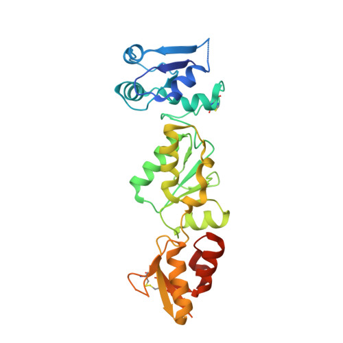
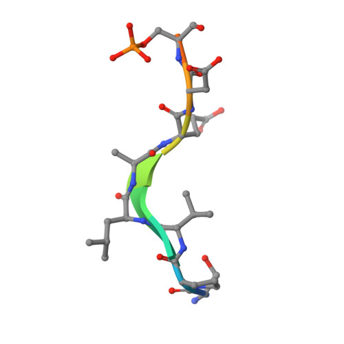
BRCT domains of the DNA damage checkpoint proteins TOPBP1/Rad4 display distinct specificities for phosphopeptide ligands.

Day, M.Rappas, M.Ptasinska, K.Boos, D.Oliver, A.W.Pearl, L.H.

(2018) Elife 7

  • DOI: https://doi.org/10.7554/eLife.39979
  • Primary Citation of Related Structures:  
    6HM3, 6HM4, 6HM5

  • PubMed Abstract: 

    TOPBP1 and its fission yeast homologueRad4, are critical players in a range of DNA replication, repair and damage signalling processes. They are composed of multiple BRCT domains, some of which bind phosphorylated motifs in other proteins. They thus act as multi-point adaptors bringing proteins together into functional combinations, dependent on post-translational modifications downstream of cell cycle and DNA damage signals. We have now structurally and/or biochemically characterised a sufficient number of high-affinity complexes for the conserved N-terminal region of TOPBP1 and Rad4 with diverse phospho-ligands, including human RAD9 and Treslin, and Schizosaccharomyces pombe Crb2 and Sld3, to define the determinants of BRCT domain specificity. We use this to identify and characterise previously unknown phosphorylation-dependent TOPBP1/Rad4-binding motifs in human RHNO1 and the fission yeast homologue of MDC1, Mdb1. These results provide important insights into how multiple BRCT domains within TOPBP1/Rad4 achieve selective and combinatorial binding of their multiple partner proteins.


  • Organizational Affiliation

    Cancer Research UK DNA Repair Enzymes Group, Genome Damage and Stability Centre, School of Life Sciences, University of Sussex, Falmer, United Kingdom.


Macromolecules
Find similar proteins by:  (by identity cutoff)  |  3D Structure
Entity ID: 1
MoleculeChains Sequence LengthOrganismDetailsImage
DNA topoisomerase II binding protein 1290Gallus gallusMutation(s): 0 
Gene Names: TOPBP1
UniProt
Find proteins for A0A1D5P3M9 (Gallus gallus)
Explore A0A1D5P3M9 
Go to UniProtKB:  A0A1D5P3M9
Entity Groups  
Sequence Clusters30% Identity50% Identity70% Identity90% Identity95% Identity100% Identity
UniProt GroupA0A1D5P3M9
Sequence Annotations
Expand
  • Reference Sequence

Find similar proteins by:  Sequence   |   3D Structure  

Entity ID: 2
MoleculeChains Sequence LengthOrganismDetailsImage
Cell cycle checkpoint control protein RAD9A11Homo sapiensMutation(s): 0 
EC: 3.1.11.2
UniProt & NIH Common Fund Data Resources
Find proteins for Q99638 (Homo sapiens)
Explore Q99638 
Go to UniProtKB:  Q99638
PHAROS:  Q99638
GTEx:  ENSG00000172613 
Entity Groups  
Sequence Clusters30% Identity50% Identity70% Identity90% Identity95% Identity100% Identity
UniProt GroupQ99638
Sequence Annotations
Expand
  • Reference Sequence
Small Molecules
Modified Residues  1 Unique
IDChains TypeFormula2D DiagramParent
SEP
Query on SEP
B
L-PEPTIDE LINKINGC3 H8 N O6 PSER
Experimental Data & Validation

Experimental Data

  • Method: X-RAY DIFFRACTION
  • Resolution: 2.33 Å
  • R-Value Free: 0.241 
  • R-Value Work: 0.201 
  • R-Value Observed: 0.205 
  • Space Group: P 1 21 1
Unit Cell:
Length ( Å )Angle ( ˚ )
a = 59.05α = 90
b = 34.07β = 103.57
c = 66.67γ = 90
Software Package:
Software NamePurpose
REFMACrefinement
PHENIXrefinement
xia2data reduction
Aimlessdata scaling
PHASERphasing

Structure Validation

View Full Validation Report


Entry History & Funding Information

Deposition Data


Funding OrganizationLocationGrant Number
Cancer Research UKUnited KingdomC302/A14532

Revision History  (Full details and data files)

  • Version 1.0: 2018-10-17
    Type: Initial release