6GYH

Crystal structure of the light-driven proton pump Coccomyxa subellipsoidea Rhodopsin CsR


Experimental Data Snapshot

  • Method: X-RAY DIFFRACTION
  • Resolution: 2.00 Å
  • R-Value Free: 0.225 
  • R-Value Work: 0.192 
  • R-Value Observed: 0.194 

wwPDB Validation   3D Report Full Report


Ligand Structure Quality Assessment 


This is version 1.2 of the entry. See complete history


Literature

Design of a light-gated proton channel based on the crystal structure ofCoccomyxarhodopsin.

Fudim, R.Szczepek, M.Vierock, J.Vogt, A.Schmidt, A.Kleinau, G.Fischer, P.Bartl, F.Scheerer, P.Hegemann, P.

(2019) Sci Signal 12

  • DOI: https://doi.org/10.1126/scisignal.aav4203
  • Primary Citation of Related Structures:  
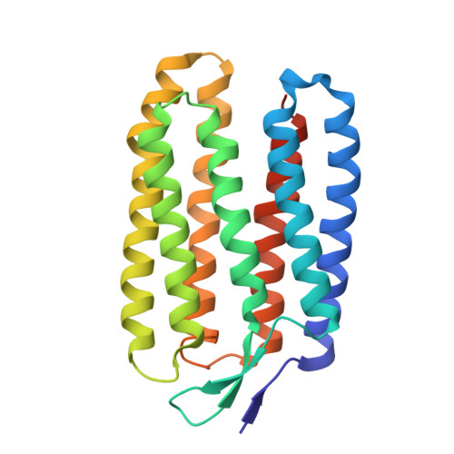
    6GYH

  • PubMed Abstract: 

    The light-driven proton pump Coccomyxa subellipsoidea rhodopsin (CsR) provides-because of its high expression in heterologous host cells-an opportunity to study active proton transport under controlled electrochemical conditions. In this study, solving crystal structure of CsR at 2.0-Å resolution enabled us to identify distinct features of the membrane protein that determine ion transport directivity and voltage sensitivity. A specific hydrogen bond between the highly conserved Arg 83 and the nearby nonconserved tyrosine (Tyr 14 ) guided our structure-based transformation of CsR into an operational light-gated proton channel (CySeR) that could potentially be used in optogenetic assays. Time-resolved electrophysiological and spectroscopic measurements distinguished pump currents from channel currents in a single protein and emphasized the necessity of Arg 83 mobility in CsR as a dynamic extracellular barrier to prevent passive conductance. Our findings reveal that molecular constraints that distinguish pump from channel currents are structurally more confined than was generally expected. This knowledge might enable the structure-based design of novel optogenetic tools, which derive from microbial pumps and are therefore ion specific.


  • Organizational Affiliation

    Experimental Biophysics, Institute for Biology, Humboldt-Universität zu Berlin, Invalidenstr. 42, 10115 Berlin, Germany.


Macromolecules
Find similar proteins by:  (by identity cutoff)  |  3D Structure
Entity ID: 1
MoleculeChains Sequence LengthOrganismDetailsImage
Family A G protein-coupled receptor-like protein236Coccomyxa subellipsoidea C-169Mutation(s): 0 
Gene Names: COCSUDRAFT_42526
Membrane Entity: Yes 
UniProt
Find proteins for I0YUS5 (Coccomyxa subellipsoidea (strain C-169))
Explore I0YUS5 
Go to UniProtKB:  I0YUS5
Entity Groups  
Sequence Clusters30% Identity50% Identity70% Identity90% Identity95% Identity100% Identity
UniProt GroupI0YUS5
Sequence Annotations
Expand
  • Reference Sequence
Experimental Data & Validation

Experimental Data

  • Method: X-RAY DIFFRACTION
  • Resolution: 2.00 Å
  • R-Value Free: 0.225 
  • R-Value Work: 0.192 
  • R-Value Observed: 0.194 
  • Space Group: H 3
Unit Cell:
Length ( Å )Angle ( ˚ )
a = 78.078α = 90
b = 78.078β = 90
c = 143.951γ = 120
Software Package:
Software NamePurpose
REFMACrefinement
XDSdata reduction
Aimlessdata scaling
PHASERphasing

Structure Validation

View Full Validation Report


Ligand Structure Quality Assessment 


Entry History & Funding Information

Deposition Data


Funding OrganizationLocationGrant Number
German Research FoundationGermanySFB1078
German Research FoundationGermanyUNICAT

Revision History  (Full details and data files)

  • Version 1.0: 2019-03-27
    Type: Initial release
  • Version 1.1: 2019-04-03
    Changes: Data collection, Database references
  • Version 1.2: 2024-01-17
    Changes: Data collection, Database references, Refinement description