6G7E

Crystal structure of Chaetomium thermophilum Mot1 (E1434Q, 1837-1886 deletion mutant)


Experimental Data Snapshot

  • Method: X-RAY DIFFRACTION
  • Resolution: 3.21 Å
  • R-Value Free: 0.238 
  • R-Value Work: 0.191 
  • R-Value Observed: 0.193 

wwPDB Validation   3D Report Full Report


This is version 1.0 of the entry. See complete history


Literature

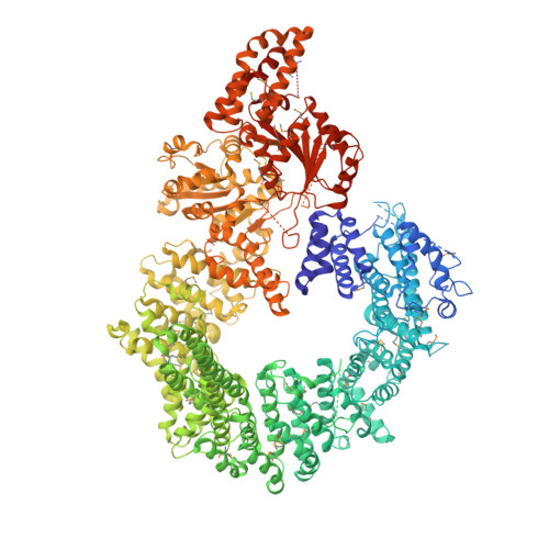
Crystal structure of the full Swi2/Snf2 remodeler Mot1 in the resting state.

Butryn, A.Woike, S.Shetty, S.J.Auble, D.T.Hopfner, K.P.

(2018) Elife 7

  • DOI: https://doi.org/10.7554/eLife.37774
  • Primary Citation of Related Structures:  
    6G7E

  • PubMed Abstract: 

    Swi2/Snf2 ATPases remodel protein:DNA complexes in all of the fundamental chromosome-associated processes. The single-subunit remodeler Mot1 dissociates TATA box-binding protein (TBP):DNA complexes and provides a simple model for obtaining structural insights into the action of Swi2/Snf2 ATPases. Previously we reported how the N-terminal domain of Mot1 binds TBP, NC2 and DNA, but the location of the C-terminal ATPase domain remained unclear (Butryn et al., 2015). Here, we report the crystal structure of the near full-length Mot1 from Chaetomium thermophilum. Our data show that Mot1 adopts a ring like structure with a catalytically inactive resting state of the ATPase. Biochemical analysis suggests that TBP binding switches Mot1 into an ATP hydrolysis-competent conformation. Combined with our previous results, these data significantly improve the structural model for the complete Mot1:TBP:DNA complex and suggest a general mechanism for Mot1 action.


  • Organizational Affiliation

    Department of Biochemistry, Ludwig-Maximilians-Universität München, Munich, Germany.


Macromolecules
Find similar proteins by:  (by identity cutoff)  |  3D Structure
Entity ID: 1
MoleculeChains Sequence LengthOrganismDetailsImage
Helicase-like proteinA [auth B]1,852Thermochaetoides thermophila DSM 1495Mutation(s): 1 
Gene Names: CTHT_0026210
UniProt
Find proteins for G0S6C0 (Chaetomium thermophilum (strain DSM 1495 / CBS 144.50 / IMI 039719))
Explore G0S6C0 
Go to UniProtKB:  G0S6C0
Entity Groups  
Sequence Clusters30% Identity50% Identity70% Identity90% Identity95% Identity100% Identity
UniProt GroupG0S6C0
Sequence Annotations
Expand
  • Reference Sequence
Small Molecules
Modified Residues  1 Unique
IDChains TypeFormula2D DiagramParent
MSE
Query on MSE
A [auth B]L-PEPTIDE LINKINGC5 H11 N O2 SeMET
Experimental Data & Validation

Experimental Data

  • Method: X-RAY DIFFRACTION
  • Resolution: 3.21 Å
  • R-Value Free: 0.238 
  • R-Value Work: 0.191 
  • R-Value Observed: 0.193 
  • Space Group: P 1 21 1
Unit Cell:
Length ( Å )Angle ( ˚ )
a = 93.134α = 90
b = 96.85β = 97.55
c = 129.64γ = 90
Software Package:
Software NamePurpose
PHENIXrefinement
XDSdata reduction
XSCALEdata scaling
PHENIXphasing

Structure Validation

View Full Validation Report


Entry History & Funding Information

Deposition Data


Funding OrganizationLocationGrant Number
European Research CouncilGermanyATMMACHINE

Revision History  (Full details and data files)

  • Version 1.0: 2018-10-17
    Type: Initial release