Molecular Insights into Function and Competitive Inhibition ofPseudomonas aeruginosaMultiple Virulence Factor Regulator.
Kitao, T., Lepine, F., Babloudi, S., Walte, F., Steinbacher, S., Maskos, K., Blaesse, M., Negri, M., Pucci, M., Zahler, B., Felici, A., Rahme, L.G.(2018) mBio 9
- PubMed: 29339431
- DOI: https://doi.org/10.1128/mBio.02158-17
- Primary Citation of Related Structures:
6B8A - PubMed Abstract:
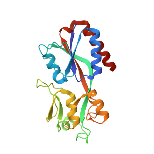
New approaches to antimicrobial drug discovery are urgently needed to combat intractable infections caused by multidrug-resistant (MDR) bacteria. M ultiple v irulence f actor r egulator (MvfR or PqsR), a Pseudomonas aeruginosa quorum sensing transcription factor, regulates functions important in both acute and persistent infections. Recently identified non-ligand-based benzamine-benzimidazole (BB) inhibitors of MvfR suppress both acute and persistent P. aeruginosa infections in mice without perturbing bacterial growth. Here, we elucidate the crystal structure of the MvfR ligand binding domain (LBD) in complex with one potent BB inhibitor, M64. Structural analysis indicated that M64 binds, like native ligands, to the MvfR hydrophobic cavity. A hydrogen bond and pi interaction were found to be important for MvfR-M64 affinity. Surface plasmon resonance analysis demonstrated that M64 is a competitive inhibitor of MvfR. Moreover, a protein engineering approach revealed that Gln194 and Tyr258 are critical for the interaction between MvfR and M64. Random mutagenesis of the full-length MvfR protein identified a single-amino-acid substitution, I68F, at a DNA binding linker domain that confers M64 insensitivity. In the presence of M64, I68F but not the wild-type (WT) MvfR protein retained DNA binding ability. Our findings strongly suggest that M64 promotes conformational change at the DNA binding domain of MvfR and that the I68F mutation may compensate for this change, indicating allosteric inhibition. This work provides critical new insights into the molecular mechanism of MvfR function and inhibition that could aid in the optimization of anti-MvfR compounds and improve our understanding of MvfR regulation. IMPORTANCE Pseudomonas aeruginosa is an opportunistic Gram-negative pathogen that causes serious acute, persistent, and relapsing infections. New approaches to antimicrobial drug discovery are urgently needed to combat intractable infections caused by this pathogen. The Pseudomonas aeruginosa quorum sensing transcription factor MvfR regulates functions important in both acute and persistent infections. We used recently identified inhibitors of MvfR to perform structural studies and reveal important insights that would benefit the optimization of anti-MvfR compounds. Altogether, the results reported here provide critical detailed mechanistic insights into the function of MvfR domains that may benefit the optimization of the chemical, pharmacological, and safety properties of MvfR antagonist series.
Organizational Affiliation:
Department of Surgery, Massachusetts General Hospital, Boston, Massachusetts, USA.