5W2A

Structure of human DNA polymerase kappa in complex with Lucidin-derived DNA adduct and incoming dCMPNPP


Experimental Data Snapshot

  • Method: X-RAY DIFFRACTION
  • Resolution: 2.90 Å
  • R-Value Free: 0.270 
  • R-Value Work: 0.231 
  • R-Value Observed: 0.233 

wwPDB Validation   3D Report Full Report


Ligand Structure Quality Assessment 


This is version 1.2 of the entry. See complete history


Literature

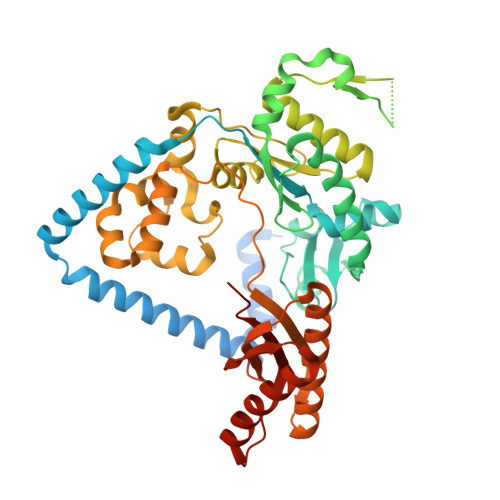
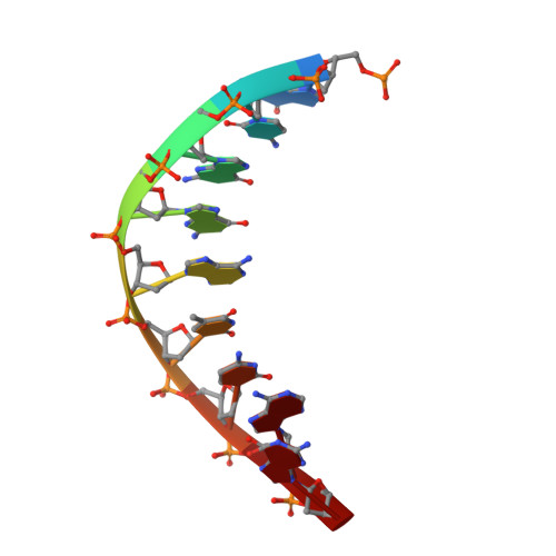
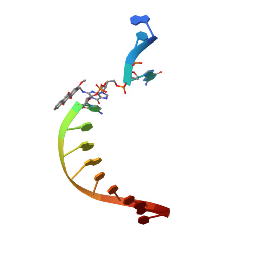
Mechanism of Error-Free DNA Replication Past Lucidin-Derived DNA Damage by Human DNA Polymerase kappa.

Yockey, O.P.Jha, V.Ghodke, P.P.Xu, T.Xu, W.Ling, H.Pradeepkumar, P.I.Zhao, L.

(2017) Chem Res Toxicol 30: 2023-2032

  • DOI: https://doi.org/10.1021/acs.chemrestox.7b00227
  • Primary Citation of Related Structures:  
    5W2A, 5W2C

  • PubMed Abstract: 

    DNA damage impinges on genetic information flow and has significant implications in human disease and aging. Lucidin-3-O-primeveroside (LuP) is an anthraquinone derivative present in madder root, which has been used as a coloring agent and food additive. LuP can be metabolically converted to genotoxic compound lucidin, which subsequently forms lucidin-specific N 2 -2'-deoxyguanosine (N 2 -dG) and N 6 -2'-deoxyadenosine (N 6 -dA) DNA adducts. Lucidin is mutagenic and carcinogenic in rodents but has low carcinogenic risks in humans. To understand the molecular mechanism of low carcinogenicity of lucidin in humans, we performed DNA replication assays using site-specifically modified oligodeoxynucleotides containing a structural analogue (LdG) of lucidin-N 2 -dG DNA adduct and determined the crystal structures of DNA polymerase (pol) κ in complex with LdG-bearing DNA and an incoming nucleotide. We examined four human pols (pol η, pol ι, pol κ, and Rev1) in their efficiency and accuracy during DNA replication with LdG; these pols are key players in translesion DNA synthesis. Our results demonstrate that pol κ efficiently and accurately replicates past the LdG adduct, whereas DNA replication by pol η, pol ι is compromised to different extents. Rev1 retains its ability to incorporate dCTP opposite the lesion albeit with decreased efficiency. Two ternary crystal structures of pol κ illustrate that the LdG adduct is accommodated by pol κ at the enzyme active site during insertion and postlesion-extension steps. The unique open active site of pol κ allows the adducted DNA to adopt a standard B-form for accurate DNA replication. Collectively, these biochemical and structural data provide mechanistic insights into the low carcinogenic risk of lucidin in humans.


  • Organizational Affiliation

    Department of Biochemistry, Schulich School of Medicine & Dentistry, University of Western Ontario , London, Ontario N6A 5C1, Canada.


Macromolecules

Find similar proteins by:  (by identity cutoff)  |  3D Structure
Entity ID: 1
MoleculeChains Sequence LengthOrganismDetailsImage
DNA polymerase kappa
A, B
551Homo sapiensMutation(s): 0 
Gene Names: POLKDINB1
EC: 2.7.7.7
UniProt & NIH Common Fund Data Resources
Find proteins for Q9UBT6 (Homo sapiens)
Explore Q9UBT6 
Go to UniProtKB:  Q9UBT6
PHAROS:  Q9UBT6
GTEx:  ENSG00000122008 
Entity Groups  
Sequence Clusters30% Identity50% Identity70% Identity90% Identity95% Identity100% Identity
UniProt GroupQ9UBT6
Sequence Annotations
Expand
  • Reference Sequence

Find similar nucleic acids by:  Sequence   |   3D Structure  

Entity ID: 2
MoleculeChains LengthOrganismImage
DNA (5'-D(P*GP*CP*GP*GP*AP*TP*CP*AP*G)-3')C,
E [auth P]
9unidentified
Sequence Annotations
Expand
  • Reference Sequence

Find similar nucleic acids by:  Sequence   |   3D Structure  

Entity ID: 3
MoleculeChains LengthOrganismImage
DNA (5'-D(*AP*TP*GP*(LDG)P*CP*TP*GP*AP*TP*CP*CP*GP*C)-3')D,
F [auth T]
13unidentified
Sequence Annotations
Expand
  • Reference Sequence
Small Molecules
Ligands 4 Unique
IDChains Name / Formula / InChI Key2D Diagram3D Interactions
0KX
Query on 0KX

Download Ideal Coordinates CCD File 
I [auth A],
N [auth B]
2'-deoxy-5'-O-[(R)-hydroxy{[(R)-hydroxy(phosphonooxy)phosphoryl]amino}phosphoryl]cytidine
C9 H17 N4 O12 P3
STYMTWKSQLVXJN-SHYZEUOFSA-N
PGE
Query on PGE

Download Ideal Coordinates CCD File 
J [auth A],
O [auth B]
TRIETHYLENE GLYCOL
C6 H14 O4
ZIBGPFATKBEMQZ-UHFFFAOYSA-N
GOL
Query on GOL

Download Ideal Coordinates CCD File 
H [auth A],
L [auth B],
M [auth B],
P
GLYCEROL
C3 H8 O3
PEDCQBHIVMGVHV-UHFFFAOYSA-N
MG
Query on MG

Download Ideal Coordinates CCD File 
G [auth A],
K [auth B]
MAGNESIUM ION
Mg
JLVVSXFLKOJNIY-UHFFFAOYSA-N
Experimental Data & Validation

Experimental Data

  • Method: X-RAY DIFFRACTION
  • Resolution: 2.90 Å
  • R-Value Free: 0.270 
  • R-Value Work: 0.231 
  • R-Value Observed: 0.233 
  • Space Group: P 21 21 21
Unit Cell:
Length ( Å )Angle ( ˚ )
a = 63.891α = 90
b = 129.014β = 90
c = 167.346γ = 90
Software Package:
Software NamePurpose
REFMACrefinement
XDSdata reduction
SCALAdata scaling
REFMACphasing

Structure Validation

View Full Validation Report


Ligand Structure Quality Assessment 


Entry History & Funding Information

Deposition Data


Funding OrganizationLocationGrant Number
Canadian Institutes of Health (CIHR)CanadaMOP-94590

Revision History  (Full details and data files)

  • Version 1.0: 2017-10-18
    Type: Initial release
  • Version 1.1: 2017-11-29
    Changes: Database references
  • Version 1.2: 2024-03-13
    Changes: Data collection, Database references, Derived calculations, Refinement description