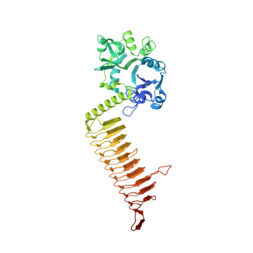
Crystal structure of a bifunctional GlmU UDP-N-acetylglucosamine diphosphorylase/glucosamine-1- phosphate N-acetyltransferase from Acinetobacter baumannii
Edwards, T.E., Abendroth, J., Lorimer, D.D., Seattle Structural Genomics Center for Infectious Disease (SSGCID)To be published.