5UOW

Triheteromeric NMDA receptor GluN1/GluN2A/GluN2B in complex with glycine, glutamate, MK-801 and a GluN2B-specific Fab, at pH 6.5


Experimental Data Snapshot

  • Method: ELECTRON MICROSCOPY
  • Resolution: 4.50 Å
  • Aggregation State: PARTICLE 
  • Reconstruction Method: SINGLE PARTICLE 

wwPDB Validation   3D Report Full Report


This is version 2.0 of the entry. See complete history


Literature

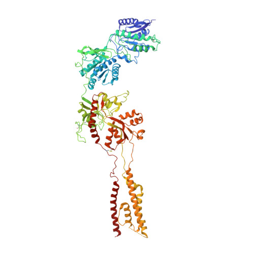
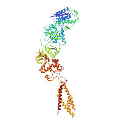
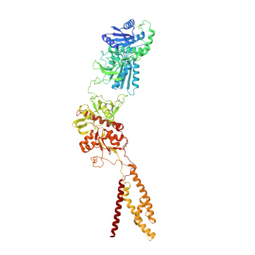
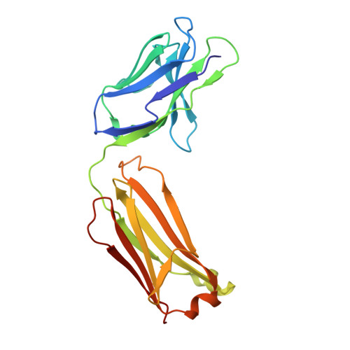
Cryo-EM structures of the triheteromeric NMDA receptor and its allosteric modulation.

Lu, W.Du, J.Goehring, A.Gouaux, E.

(2017) Science 355

  • DOI: https://doi.org/10.1126/science.aal3729
  • Primary Citation of Related Structures:  
    5UOW, 5UP2

  • PubMed Abstract: 

    N -methyl-d-aspartate receptors (NMDARs) are heterotetrameric ion channels assembled as diheteromeric or triheteromeric complexes. Here, we report structures of the triheteromeric GluN1/GluN2A/GluN2B receptor in the absence or presence of the GluN2B-specific allosteric modulator Ro 25-6981 (Ro), determined by cryogenic electron microscopy (cryo-EM). In the absence of Ro, the GluN2A and GluN2B amino-terminal domains (ATDs) adopt "closed" and "open" clefts, respectively. Upon binding Ro, the GluN2B ATD clamshell transitions from an open to a closed conformation. Consistent with a predominance of the GluN2A subunit in ion channel gating, the GluN2A subunit interacts more extensively with GluN1 subunits throughout the receptor, in comparison with the GluN2B subunit. Differences in the conformation of the pseudo-2-fold-related GluN1 subunits further reflect receptor asymmetry. The triheteromeric NMDAR structures provide the first view of the most common NMDA receptor assembly and show how incorporation of two different GluN2 subunits modifies receptor symmetry and subunit interactions, allowing each subunit to uniquely influence receptor structure and function, thus increasing receptor complexity.


  • Organizational Affiliation

    Vollum Institute, Oregon Health and Science University, 3181 Southwest Sam Jackson Park Road, Portland, OR 97239, USA.


Macromolecules
Find similar proteins by:  (by identity cutoff)  |  3D Structure
Entity ID: 1
MoleculeChains Sequence LengthOrganismDetailsImage
N-methyl-D-aspartate receptor subunit NR1-8a
A, C
814Xenopus laevisMutation(s): 0 
Membrane Entity: Yes 
UniProt
Find proteins for A0A1L8F5J9 (Xenopus laevis)
Explore A0A1L8F5J9 
Go to UniProtKB:  A0A1L8F5J9
Entity Groups  
Sequence Clusters30% Identity50% Identity70% Identity90% Identity95% Identity100% Identity
UniProt GroupA0A1L8F5J9
Sequence Annotations
Expand
  • Reference Sequence
Find similar proteins by:  (by identity cutoff)  |  3D Structure
Entity ID: 2
MoleculeChains Sequence LengthOrganismDetailsImage
N-methyl-D-aspartate receptor subunit NR2A832Xenopus laevisMutation(s): 0 
Gene Names: LOC100127346
Membrane Entity: Yes 
UniProt
Find proteins for B7ZSK1 (Xenopus laevis)
Explore B7ZSK1 
Go to UniProtKB:  B7ZSK1
Entity Groups  
Sequence Clusters30% Identity50% Identity70% Identity90% Identity95% Identity100% Identity
UniProt GroupB7ZSK1
Sequence Annotations
Expand
  • Reference Sequence
Find similar proteins by:  (by identity cutoff)  |  3D Structure
Entity ID: 3
MoleculeChains Sequence LengthOrganismDetailsImage
Ionotropic glutamate receptor subunit NR2B837Xenopus laevisMutation(s): 0 
Gene Names: NR2B
Membrane Entity: Yes 
UniProt
Find proteins for A7XY94 (Xenopus laevis)
Explore A7XY94 
Go to UniProtKB:  A7XY94
Entity Groups  
Sequence Clusters30% Identity50% Identity70% Identity90% Identity95% Identity100% Identity
UniProt GroupA7XY94
Sequence Annotations
Expand
  • Reference Sequence
Find similar proteins by:  (by identity cutoff)  |  3D Structure
Entity ID: 4
MoleculeChains Sequence LengthOrganismDetailsImage
GluN2B-specific Fab, termed 11D1E [auth F],
F [auth G]
216Mus musculusMutation(s): 0 
Sequence Annotations
Expand
  • Reference Sequence
Oligosaccharides

Help

Entity ID: 5
MoleculeChains Length2D Diagram Glycosylation3D Interactions
2-acetamido-2-deoxy-beta-D-glucopyranose-(1-4)-2-acetamido-2-deoxy-beta-D-glucopyranose
G [auth E],
H,
I,
J,
K,
G [auth E],
H,
I,
J,
K,
L,
M,
N
2N-Glycosylation
Glycosylation Resources
GlyTouCan:  G42666HT
GlyCosmos:  G42666HT
GlyGen:  G42666HT
Small Molecules
Ligands 2 Unique
IDChains Name / Formula / InChI Key2D Diagram3D Interactions
BMK
Query on BMK

Download Ideal Coordinates CCD File 
P [auth B](5S,10R)-5-methyl-10,11-dihydro-5H-5,10-epiminodibenzo[a,d][7]annulene
C16 H15 N
LBOJYSIDWZQNJS-CVEARBPZSA-N
GLU
Query on GLU

Download Ideal Coordinates CCD File 
O [auth B]GLUTAMIC ACID
C5 H9 N O4
WHUUTDBJXJRKMK-VKHMYHEASA-N
Binding Affinity Annotations 
IDSourceBinding Affinity
BMK BindingDB:  5UOW Ki: min: 8, max: 31 (nM) from 3 assay(s)
Experimental Data & Validation

Experimental Data

  • Method: ELECTRON MICROSCOPY
  • Resolution: 4.50 Å
  • Aggregation State: PARTICLE 
  • Reconstruction Method: SINGLE PARTICLE 

Structure Validation

View Full Validation Report


Entry History 

Deposition Data

Revision History  (Full details and data files)

  • Version 1.0: 2017-03-22
    Type: Initial release
  • Version 1.1: 2017-11-08
    Changes: Advisory, Data collection, Derived calculations
  • Version 1.2: 2018-10-24
    Changes: Data collection, Database references, Structure summary
  • Version 2.0: 2020-07-29
    Type: Remediation
    Reason: Carbohydrate remediation
    Changes: Advisory, Atomic model, Data collection, Derived calculations, Structure summary