5UI2

CRYSTAL STRUCTURE OF ORANGE CAROTENOID PROTEIN


Experimental Data Snapshot

  • Method: X-RAY DIFFRACTION
  • Resolution: 2.10 Å
  • R-Value Free: 0.266 
  • R-Value Work: 0.213 
  • R-Value Observed: 0.215 

wwPDB Validation   3D Report Full Report


Ligand Structure Quality Assessment 


This is version 2.1 of the entry. See complete history


Literature

The crystal structure of a cyanobacterial water-soluble carotenoid binding protein.

Kerfeld, C.A.Sawaya, M.R.Brahmandam, V.Cascio, D.Ho, K.K.Trevithick-Sutton, C.C.Krogmann, D.W.Yeates, T.O.

(2003) Structure 11: 55-65

  • DOI: https://doi.org/10.1016/s0969-2126(02)00936-x
  • Primary Citation of Related Structures:  
    5UI2

  • PubMed Abstract: 

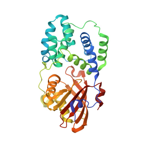
    Carotenoids undergo a wide range of photochemical reactions in animal, plant, and microbial systems. In photosynthetic organisms, in addition to light harvesting, they perform an essential role in protecting against light-induced damage by quenching singlet oxygen, superoxide anion radicals, or triplet-state chlorophyll. We have determined the crystal structure of a water-soluble orange carotenoid protein (OCP) isolated from the cyanobacterium Arthrospira maxima at a resolution of 2.1 A. OCP forms a homodimer with one carotenoid molecule per monomer. The carotenoid binding site is lined by a striking number of methionine residues. The structure reveals several possible ways in which the protein environment influences the spectral properties of the pigment and provides insight into how the OCP carries out its putative functions in photoprotection.


  • Organizational Affiliation

    Molecular Biology Institute, University of California, Los Angeles, Los Angeles, CA 90095, USA. kerfeld@mbi.ucla.edu


Macromolecules
Find similar proteins by:  (by identity cutoff)  |  3D Structure
Entity ID: 1
MoleculeChains Sequence LengthOrganismDetailsImage
Orange carotenoid-binding protein
A, B
317Limnospira maximaMutation(s): 0 
UniProt
Find proteins for P83689 (Limnospira maxima)
Explore P83689 
Go to UniProtKB:  P83689
Entity Groups  
Sequence Clusters30% Identity50% Identity70% Identity90% Identity95% Identity100% Identity
UniProt GroupP83689
Sequence Annotations
Expand
  • Reference Sequence
Oligosaccharides

Help

Entity ID: 2
MoleculeChains Length2D Diagram Glycosylation3D Interactions
beta-D-fructofuranose-(2-1)-alpha-D-glucopyranose
C
2N/A
Glycosylation Resources
GlyTouCan:  G05551OP
GlyCosmos:  G05551OP
Biologically Interesting Molecules (External Reference) 1 Unique
Experimental Data & Validation

Experimental Data

  • Method: X-RAY DIFFRACTION
  • Resolution: 2.10 Å
  • R-Value Free: 0.266 
  • R-Value Work: 0.213 
  • R-Value Observed: 0.215 
  • Space Group: C 1 2 1
Unit Cell:
Length ( Å )Angle ( ˚ )
a = 217.2α = 90
b = 40.761β = 95.8
c = 75.46γ = 90
Software Package:
Software NamePurpose
BUSTERrefinement
PDB_EXTRACTdata extraction
DENZOdata reduction
SCALEPACKdata scaling
MLPHAREphasing

Structure Validation

View Full Validation Report


Ligand Structure Quality Assessment 


Entry History & Funding Information

Deposition Data


Funding OrganizationLocationGrant Number
National Institutes of Health/National Institute of General Medical Sciences (NIH/NIGMS)United StatesNIH GM 31299
United States Department of Agriculture (USDA)United StatesUSDA1999-01759

Revision History  (Full details and data files)

  • Version 1.0: 2017-01-25
    Type: Initial release
  • Version 1.1: 2017-09-27
    Changes: Author supporting evidence
  • Version 1.2: 2018-10-10
    Changes: Data collection, Database references, Structure summary
  • Version 1.3: 2020-01-01
    Changes: Author supporting evidence, Structure summary
  • Version 2.0: 2020-07-29
    Type: Remediation
    Reason: Carbohydrate remediation
    Changes: Atomic model, Data collection, Derived calculations, Non-polymer description, Structure summary
  • Version 2.1: 2024-03-06
    Changes: Data collection, Database references, Structure summary