5T3K

Fluorescence detection of RNA-ligand binding and crystal structure determination of ribosomal decoding site RNA using a heavy atom containing fluorescent ribonucleoside


Experimental Data Snapshot

  • Method: X-RAY DIFFRACTION
  • Resolution: 2.14 Å
  • R-Value Free: 0.246 
  • R-Value Work: 0.197 
  • R-Value Observed: 0.203 

wwPDB Validation   3D Report Full Report


This is version 1.1 of the entry. See complete history


Literature

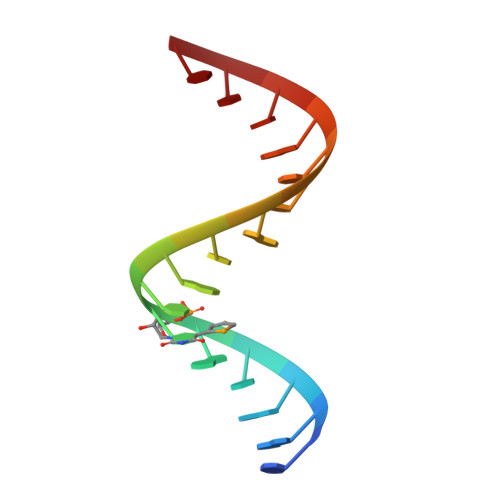
Structure of the Ribosomal RNA Decoding Site Containing a Selenium-Modified Responsive Fluorescent Ribonucleoside Probe.

Nuthanakanti, A.Boerneke, M.A.Hermann, T.Srivatsan, S.G.

(2017) Angew Chem Int Ed Engl 56: 2640-2644

  • DOI: https://doi.org/10.1002/anie.201611700
  • Primary Citation of Related Structures:  
    5T3K

  • PubMed Abstract: 

    Comprehensive understanding of the structure-function relationship of RNA both in real time and at atomic level will have a profound impact in advancing our understanding of RNA functions in biology. Here, we describe the first example of a multifunctional nucleoside probe, containing a conformation-sensitive fluorophore and an anomalous X-ray diffraction label (5-selenophene uracil), which enables the correlation of RNA conformation and recognition under equilibrium and in 3D. The probe incorporated into the bacterial ribosomal RNA decoding site, fluorescently reports antibiotic binding and provides diffraction information in determining the structure without distorting native RNA fold. Further, by comparing solution binding data and crystal structure, we gained insight on how the probe senses ligand-induced conformational change in RNA. Taken together, our nucleoside probe represents a new class of biophysical tool that would complement available tools for functional RNA investigations.


  • Organizational Affiliation

    Department of Chemistry, Indian Institute of Science Education and Research, Dr. Homi Bhabha Road, Pashan, Pune, 411008, India.


Macromolecules

Find similar nucleic acids by:  Sequence   |   3D Structure  

Entity ID: 1
MoleculeChains LengthOrganismImage
RNA (5'-R(*GP*GP*UP*GP*GP*UP*GP*AP*AP*GP*UP*CP*GP*CP*UP*GP*G)-3')
A, C
17synthetic construct
Sequence Annotations
Expand
  • Reference Sequence

Find similar nucleic acids by:  Sequence   |   3D Structure  

Entity ID: 2
MoleculeChains LengthOrganismImage
RNA (5'-R(*CP*AP*GP*CP*GP*(75B)P*CP*AP*CP*AP*CP*CP*AP*CP*CP*C)-3')
B, D
16synthetic construct
Sequence Annotations
Expand
  • Reference Sequence
Experimental Data & Validation

Experimental Data

  • Method: X-RAY DIFFRACTION
  • Resolution: 2.14 Å
  • R-Value Free: 0.246 
  • R-Value Work: 0.197 
  • R-Value Observed: 0.203 
  • Space Group: P 1 21 1
Unit Cell:
Length ( Å )Angle ( ˚ )
a = 31.553α = 90
b = 86.956β = 94.14
c = 32.564γ = 90
Software Package:
Software NamePurpose
PHENIXrefinement
HKL-2000data reduction
HKL-2000data scaling

Structure Validation

View Full Validation Report


Entry History 

Deposition Data

Revision History  (Full details and data files)

  • Version 1.0: 2017-07-05
    Type: Initial release
  • Version 1.1: 2024-03-06
    Changes: Data collection, Database references, Derived calculations