5T2O

Engineered variant of I-OnuI meganuclease targeting the Anopheles AGAP011377 gene; harbors 53 point mutations relative to wild-type I-OnuI


Experimental Data Snapshot

  • Method: X-RAY DIFFRACTION
  • Resolution: 2.80 Å
  • R-Value Free: 0.281 
  • R-Value Work: 0.224 
  • R-Value Observed: 0.230 

wwPDB Validation   3D Report Full Report


This is version 1.4 of the entry. See complete history


Literature

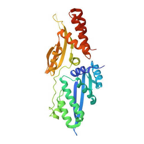
Crystallographic analyses illustrate significant plasticity and efficient recoding of meganuclease target specificity.

Werther, R.Hallinan, J.P.Lambert, A.R.Havens, K.Pogson, M.Jarjour, J.Galizi, R.Windbichler, N.Crisanti, A.Nolan, T.Stoddard, B.L.

(2017) Nucleic Acids Res 45: 8621-8634

  • DOI: https://doi.org/10.1093/nar/gkx544
  • Primary Citation of Related Structures:  
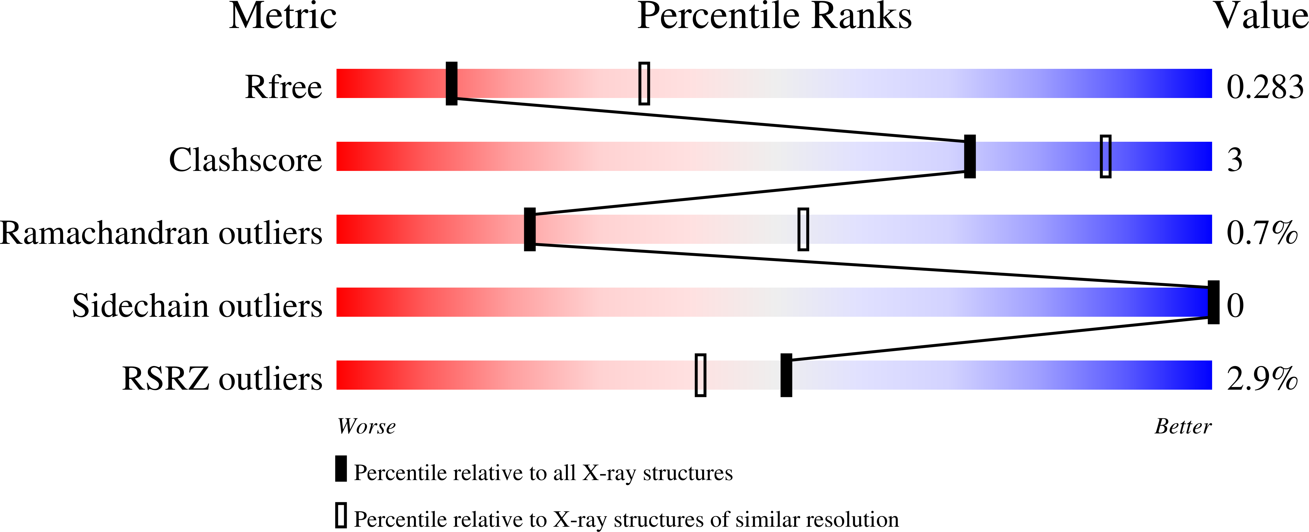
    5T2H, 5T2N, 5T2O

  • PubMed Abstract: 

    The retargeting of protein-DNA specificity, outside of extremely modular DNA binding proteins such as TAL effectors, has generally proved to be quite challenging. Here, we describe structural analyses of five different extensively retargeted variants of a single homing endonuclease, that have been shown to function efficiently in ex vivo and in vivo applications. The redesigned proteins harbor mutations at up to 53 residues (18%) of their amino acid sequence, primarily distributed across the DNA binding surface, making them among the most significantly reengineered ligand-binding proteins to date. Specificity is derived from the combined contributions of DNA-contacting residues and of neighboring residues that influence local structural organization. Changes in specificity are facilitated by the ability of all those residues to readily exchange both form and function. The fidelity of recognition is not precisely correlated with the fraction or total number of residues in the protein-DNA interface that are actually involved in DNA contacts, including directional hydrogen bonds. The plasticity of the DNA-recognition surface of this protein, which allows substantial retargeting of recognition specificity without requiring significant alteration of the surrounding protein architecture, reflects the ability of the corresponding genetic elements to maintain mobility and persistence in the face of genetic drift within potential host target sites.


  • Organizational Affiliation

    Basic Sciences Division, Fred Hutchinson Cancer Research Center, 1100 Fairview Ave. N., Seattle, WA 98109, USA.


Macromolecules

Find similar proteins by:  (by identity cutoff)  |  3D Structure
Entity ID: 1
MoleculeChains Sequence LengthOrganismDetailsImage
I-OnuI_e-ag011377302synthetic constructMutation(s): 0 
Entity Groups  
Sequence Clusters30% Identity50% Identity70% Identity90% Identity95% Identity100% Identity
Sequence Annotations
Expand
  • Reference Sequence
Find similar nucleic acids by:  (by identity cutoff)  |  3D Structure
Entity ID: 2
MoleculeChains LengthOrganismImage
DNA (26-MER)26synthetic construct
Sequence Annotations
Expand
  • Reference Sequence
Find similar nucleic acids by:  (by identity cutoff)  |  3D Structure
Entity ID: 3
MoleculeChains LengthOrganismImage
DNA (26-MER)26synthetic construct
Sequence Annotations
Expand
  • Reference Sequence
Experimental Data & Validation

Experimental Data

  • Method: X-RAY DIFFRACTION
  • Resolution: 2.80 Å
  • R-Value Free: 0.281 
  • R-Value Work: 0.224 
  • R-Value Observed: 0.230 
  • Space Group: P 21 21 21
Unit Cell:
Length ( Å )Angle ( ˚ )
a = 40.18α = 90
b = 64.835β = 90
c = 167.005γ = 90
Software Package:
Software NamePurpose
PHENIXrefinement
HKL-2000data scaling
PHASERphasing
PDB_EXTRACTdata extraction
HKL-2000data reduction

Structure Validation

View Full Validation Report


Entry History & Funding Information

Deposition Data


Funding OrganizationLocationGrant Number
National Institutes of Health/National Institute of General Medical Sciences (NIH/NIGMS)United StatesR01 GM105691
Bill & Melinda Gates FoundationUnited States--
bluebird bio, inc. (Corporate Funding)United States--

Revision History  (Full details and data files)

  • Version 1.0: 2017-05-03
    Type: Initial release
  • Version 1.1: 2017-09-27
    Changes: Author supporting evidence
  • Version 1.2: 2019-12-25
    Changes: Author supporting evidence
  • Version 1.3: 2021-06-23
    Changes: Database references, Derived calculations
  • Version 1.4: 2023-10-04
    Changes: Data collection, Database references, Refinement description