5OVQ

Crystal Structure of the peroxiredoxin (AhpC2) from the Hyperthermophilic bacteria Aquifex aeolicus VF


Experimental Data Snapshot

  • Method: X-RAY DIFFRACTION
  • Resolution: 1.80 Å
  • R-Value Free: 0.214 
  • R-Value Work: 0.185 
  • R-Value Observed: 0.187 

wwPDB Validation   3D Report Full Report


This is version 1.1 of the entry. See complete history


Literature

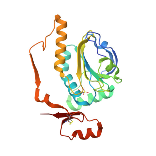
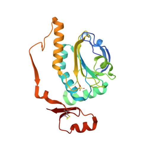
Structural properties of the peroxiredoxin AhpC2 from the hyperthermophilic eubacterium Aquifex aeolicus.

Liu, W.Liu, A.Gao, H.Wang, Q.Wang, L.Warkentin, E.Rao, Z.Michel, H.Peng, G.

(2018) Biochim Biophys Acta Gen Subj 1862: 2797-2805

  • DOI: https://doi.org/10.1016/j.bbagen.2018.08.017
  • Primary Citation of Related Structures:  
    5OVQ

  • PubMed Abstract: 

    Peroxiredoxins (Prxs) are thiol peroxidases that scavenge various peroxide substrates such as hydrogen peroxide (H 2 O 2 ), alkyl hydroperoxides and peroxinitrite. They also function as chaperones and are involved in signal transduction by H 2 O 2 in eukaryotic cells. The genome of Aquifex aeolicus, a microaerophilic, hyperthermophilic eubacterium, encodes four Prxs, among them an alkyl hydroperoxide reductase AhpC2 which was found to be closely related to archaeal 1-Cys peroxiredoxins. We determined the crystal structure of AhpC2 at 1.8 Å resolution and investigated its oligomeric state in solution by electron microscopy. AhpC2 is arranged as a toroid-shaped dodecamer instead of the typically observed decamer. The basic folding topology and the active site structure are conserved and possess a high structural similarity to other 1-Cys Prxs. However, the C-terminal region adopts an opposite orientation. AhpC2 contains three cysteines, Cys 49 , Cys 212 , and Cys 218 . The peroxidatic cysteine C P 49 was found to be hyperoxidized to the sulfonic acid (SO 3 H) form, while Cys 212 forms an intra-monomer disulfide bond with Cys 218 . Mutagenesis experiments indicate that Cys 212 and Cys 218 play important roles in the oligomerization of AhpC2. Based on these structural characteristics, we proposed the catalytic mechanism of AhpC2. This study provides novel insights into the structure and reaction mechanism of 1-Cys peroxiredoxins.


  • Organizational Affiliation

    Department of Molecular Membrane Biology, Max Planck Institute of Biophysics, 60438 Frankfurt am Main, Germany.


Macromolecules
Find similar proteins by:  (by identity cutoff)  |  3D Structure
Entity ID: 1
MoleculeChains Sequence LengthOrganismDetailsImage
Peroxiredoxin
A, B, C, E, K
223Aquifex aeolicus VF5Mutation(s): 0 
EC: 1.11.1.15
UniProt
Find proteins for O67024 (Aquifex aeolicus (strain VF5))
Explore O67024 
Go to UniProtKB:  O67024
Entity Groups  
Sequence Clusters30% Identity50% Identity70% Identity90% Identity95% Identity100% Identity
UniProt GroupO67024
Sequence Annotations
Expand
  • Reference Sequence
Find similar proteins by:  (by identity cutoff)  |  3D Structure
Entity ID: 2
MoleculeChains Sequence LengthOrganismDetailsImage
Peroxiredoxin
D, F, G, H, I
D, F, G, H, I, J, L
222Aquifex aeolicus VF5Mutation(s): 0 
EC: 1.11.1.15
UniProt
Find proteins for O67024 (Aquifex aeolicus (strain VF5))
Explore O67024 
Go to UniProtKB:  O67024
Entity Groups  
Sequence Clusters30% Identity50% Identity70% Identity90% Identity95% Identity100% Identity
UniProt GroupO67024
Sequence Annotations
Expand
  • Reference Sequence
Experimental Data & Validation

Experimental Data

  • Method: X-RAY DIFFRACTION
  • Resolution: 1.80 Å
  • R-Value Free: 0.214 
  • R-Value Work: 0.185 
  • R-Value Observed: 0.187 
  • Space Group: P 1
Unit Cell:
Length ( Å )Angle ( ˚ )
a = 51.1α = 61
b = 141.5β = 79.4
c = 143γ = 80.9
Software Package:
Software NamePurpose
PHENIXrefinement
XDSdata reduction
XDSdata scaling
PHASERphasing

Structure Validation

View Full Validation Report


Entry History 

Deposition Data

Revision History  (Full details and data files)

  • Version 1.0: 2018-10-10
    Type: Initial release
  • Version 1.1: 2024-01-17
    Changes: Data collection, Database references, Refinement description