5O63

Crystal structure of UbaLAI restriction endonuclease B3 domain domain (mutant L24M L53M L95M) with cognate DNA


Experimental Data Snapshot

  • Method: X-RAY DIFFRACTION
  • Resolution: 1.60 Å
  • R-Value Free: 0.191 
  • R-Value Work: 0.159 
  • R-Value Observed: 0.162 

wwPDB Validation   3D Report Full Report


This is version 1.1 of the entry. See complete history


Literature

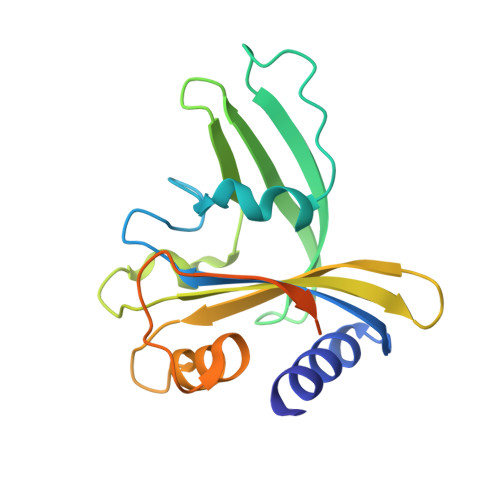
UbaLAI is a monomeric Type IIE restriction enzyme.

Sasnauskas, G.Tamulaitiene, G.Tamulaitis, G.Calyseva, J.Laime, M.Rimseliene, R.Lubys, A.Siksnys, V.

(2017) Nucleic Acids Res 45: 9583-9594

  • DOI: https://doi.org/10.1093/nar/gkx634
  • Primary Citation of Related Structures:  
    5O63

  • PubMed Abstract: 

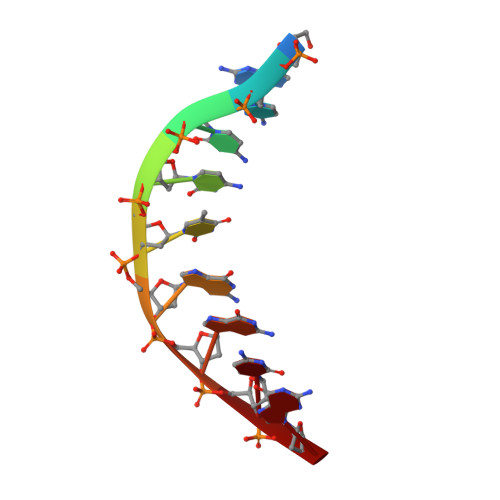
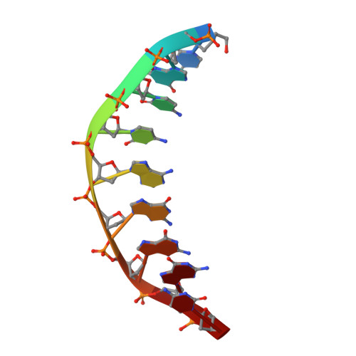
    Type II restriction endonucleases (REases) form a large and highly diverse group of enzymes. Even REases specific for a common recognition site often vary in their oligomeric structure, domain organization and DNA cleavage mechanisms. Here we report biochemical and structural characterization of the monomeric restriction endonuclease UbaLAI, specific for the pseudosymmetric DNA sequence 5'-CC/WGG-3' (where W = A/T, and '/' marks the cleavage position). We present a 1.6 Å co-crystal structure of UbaLAI N-terminal domain (UbaLAI-N) and show that it resembles the B3-family domain of EcoRII specific for the 5'-CCWGG-3' sequence. We also find that UbaLAI C-terminal domain (UbaLAI-C) is closely related to the monomeric REase MvaI, another enzyme specific for the 5'-CCWGG-3' sequence. Kinetic studies of UbaLAI revealed that it requires two recognition sites for optimal activity, and, like other type IIE enzymes, uses one copy of a recognition site to stimulate cleavage of a second copy. We propose that during the reaction UbaLAI-N acts as a handle that tethers the monomeric UbaLAI-C domain to the DNA, thereby helping UbaLAI-C to perform two sequential DNA nicking reactions on the second recognition site during a single DNA-binding event. A similar reaction mechanism may be characteristic to other monomeric two-domain REases.


  • Organizational Affiliation

    Institute of Biotechnology, Vilnius University, Sauletekio av. 7, LT-10257 Vilnius, Lithuania.


Macromolecules

Find similar proteins by:  (by identity cutoff)  |  3D Structure
Entity ID: 1
MoleculeChains Sequence LengthOrganismDetailsImage
Restriction endonuclease UbaLAI
A, B
186unidentifiedMutation(s): 0 
EC: 3.1.21.4
Entity Groups  
Sequence Clusters30% Identity50% Identity70% Identity90% Identity95% Identity100% Identity
Sequence Annotations
Expand
  • Reference Sequence

Find similar nucleic acids by:  Sequence   |   3D Structure  

Entity ID: 2
MoleculeChains LengthOrganismImage
DNA (5'-D(*C*G*C*C*A*G*G*G*C*)-3')
C, E
9synthetic construct
Sequence Annotations
Expand
  • Reference Sequence

Find similar nucleic acids by:  Sequence   |   3D Structure  

Entity ID: 3
MoleculeChains LengthOrganismImage
DNA (5'-D(*G*C*C*C*T*G*G*C*G*)-3')
D, F
9synthetic construct
Sequence Annotations
Expand
  • Reference Sequence
Experimental Data & Validation

Experimental Data

  • Method: X-RAY DIFFRACTION
  • Resolution: 1.60 Å
  • R-Value Free: 0.191 
  • R-Value Work: 0.159 
  • R-Value Observed: 0.162 
  • Space Group: P 21 21 21
Unit Cell:
Length ( Å )Angle ( ˚ )
a = 40.775α = 90
b = 94.583β = 90
c = 123.403γ = 90
Software Package:
Software NamePurpose
SCALAdata scaling
SHELXphasing
Cootmodel building
PHENIXrefinement
PDB_EXTRACTdata extraction
XDSdata reduction

Structure Validation

View Full Validation Report


Entry History & Funding Information

Deposition Data


Funding OrganizationLocationGrant Number
Research Council of LithuaniaLithuaniaMIP-81/2010

Revision History  (Full details and data files)

  • Version 1.0: 2017-08-02
    Type: Initial release
  • Version 1.1: 2017-10-04
    Changes: Database references