5N69

Cardiac muscle myosin S1 fragment in the pre-powerstroke state co-crystallized with the activator Omecamtiv Mecarbil


Experimental Data Snapshot

  • Method: X-RAY DIFFRACTION
  • Resolution: 2.45 Å
  • R-Value Free: 0.224 
  • R-Value Work: 0.180 
  • R-Value Observed: 0.182 

wwPDB Validation   3D Report Full Report


Ligand Structure Quality Assessment 


This is version 1.2 of the entry. See complete history

Re-refinement Note

A newer entry is available that reflects an alternative modeling of the original data: 8QYU


Literature

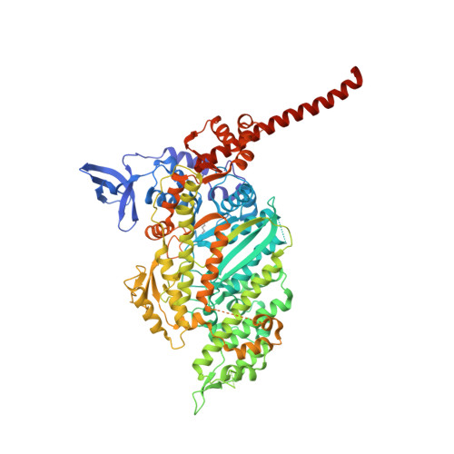
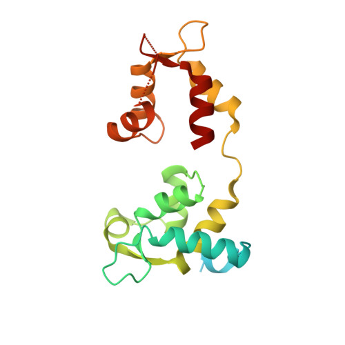
Mechanistic and structural basis for activation of cardiac myosin force production by omecamtiv mecarbil.

Planelles-Herrero, V.J.Hartman, J.J.Robert-Paganin, J.Malik, F.I.Houdusse, A.

(2017) Nat Commun 8: 190-190

  • DOI: https://doi.org/10.1038/s41467-017-00176-5
  • Primary Citation of Related Structures:  
    5N69, 5N6A

  • PubMed Abstract: 

    Omecamtiv mecarbil is a selective, small-molecule activator of cardiac myosin that is being developed as a potential treatment for heart failure with reduced ejection fraction. Here we determine the crystal structure of cardiac myosin in the pre-powerstroke state, the most relevant state suggested by kinetic studies, both with (2.45 Å) and without (3.10 Å) omecamtiv mecarbil bound. Omecamtiv mecarbil does not change the motor mechanism nor does it influence myosin structure. Instead, omecamtiv mecarbil binds to an allosteric site that stabilizes the lever arm in a primed position resulting in accumulation of cardiac myosin in the primed state prior to onset of cardiac contraction, thus increasing the number of heads that can bind to the actin filament and undergo a powerstroke once the cardiac cycle starts. The mechanism of action of omecamtiv mecarbil also provides insights into uncovering how force is generated by molecular motors.Omecamtiv mecarbil (OM) is a cardiac myosin activator that is currently in clinical trials for heart failure treatment. Here, the authors give insights into its mode of action and present the crystal structure of OM bound to bovine cardiac myosin, which shows that OM stabilizes the pre-powerstroke state of myosin.


  • Organizational Affiliation

    Structural Motility, Institut Curie, PSL Research University, CNRS, UMR 144, F-75005, Paris, France.


Macromolecules
Find similar proteins by:  (by identity cutoff)  |  3D Structure
Entity ID: 1
MoleculeChains Sequence LengthOrganismDetailsImage
Myosin-7
A, B
828Bos taurusMutation(s): 0 
UniProt
Find proteins for Q9BE39 (Bos taurus)
Explore Q9BE39 
Go to UniProtKB:  Q9BE39
Entity Groups  
Sequence Clusters30% Identity50% Identity70% Identity90% Identity95% Identity100% Identity
UniProt GroupQ9BE39
Sequence Annotations
Expand
  • Reference Sequence
Find similar proteins by:  (by identity cutoff)  |  3D Structure
Entity ID: 2
MoleculeChains Sequence LengthOrganismDetailsImage
Myosin light chain 3C [auth G],
D [auth H]
199Bos taurusMutation(s): 0 
UniProt
Find proteins for P85100 (Bos taurus)
Explore P85100 
Go to UniProtKB:  P85100
Entity Groups  
Sequence Clusters30% Identity50% Identity70% Identity90% Identity95% Identity100% Identity
UniProt GroupP85100
Sequence Annotations
Expand
  • Reference Sequence
Small Molecules
Ligands 6 Unique
IDChains Name / Formula / InChI Key2D Diagram3D Interactions
ADP
Query on ADP

Download Ideal Coordinates CCD File 
E [auth A],
V [auth B]
ADENOSINE-5'-DIPHOSPHATE
C10 H15 N5 O10 P2
XTWYTFMLZFPYCI-KQYNXXCUSA-N
2OW
Query on 2OW

Download Ideal Coordinates CCD File 
H [auth A],
Y [auth B]
methyl 4-(2-fluoro-3-{[(6-methylpyridin-3-yl)carbamoyl]amino}benzyl)piperazine-1-carboxylate
C20 H24 F N5 O3
RFUBTTPMWSKEIW-UHFFFAOYSA-N
TCE
Query on TCE

Download Ideal Coordinates CCD File 
IA [auth B]3,3',3''-phosphanetriyltripropanoic acid
C9 H15 O6 P
PZBFGYYEXUXCOF-UHFFFAOYSA-N
VO4
Query on VO4

Download Ideal Coordinates CCD File 
F [auth A],
W [auth B]
VANADATE ION
O4 V
LSGOVYNHVSXFFJ-UHFFFAOYSA-N
GOL
Query on GOL

Download Ideal Coordinates CCD File 
AA [auth B]
BA [auth B]
CA [auth B]
DA [auth B]
EA [auth B]
AA [auth B],
BA [auth B],
CA [auth B],
DA [auth B],
EA [auth B],
FA [auth B],
GA [auth B],
HA [auth B],
I [auth A],
J [auth A],
JA [auth H],
K [auth A],
KA [auth H],
L [auth A],
LA [auth H],
M [auth A],
N [auth A],
O [auth A],
P [auth A],
Q [auth A],
R [auth A],
S [auth A],
T [auth A],
U [auth A],
Z [auth B]
GLYCEROL
C3 H8 O3
PEDCQBHIVMGVHV-UHFFFAOYSA-N
MG
Query on MG

Download Ideal Coordinates CCD File 
G [auth A],
X [auth B]
MAGNESIUM ION
Mg
JLVVSXFLKOJNIY-UHFFFAOYSA-N
Modified Residues  1 Unique
IDChains TypeFormula2D DiagramParent
M3L
Query on M3L
A, B
L-PEPTIDE LINKINGC9 H21 N2 O2LYS
Binding Affinity Annotations 
IDSourceBinding Affinity
2OW Binding MOAD:  5N69 Kd: 290 (nM) from 1 assay(s)
Experimental Data & Validation

Experimental Data

  • Method: X-RAY DIFFRACTION
  • Resolution: 2.45 Å
  • R-Value Free: 0.224 
  • R-Value Work: 0.180 
  • R-Value Observed: 0.182 
  • Space Group: P 21 21 21
Unit Cell:
Length ( Å )Angle ( ˚ )
a = 98.32α = 90
b = 122.45β = 90
c = 187.43γ = 90
Software Package:
Software NamePurpose
BUSTERrefinement
XDSdata reduction
XDSdata scaling
MOLREPphasing

Structure Validation

View Full Validation Report


Ligand Structure Quality Assessment 


Entry History & Funding Information

Deposition Data


Funding OrganizationLocationGrant Number
AFMFrance18423
FRMFranceDBI20141231319
French National Research AgencyFrance13-BSV8-0019-01
AFMFrance17235
Labex CelTisPhyBioFrance11-LBX-0038
IDEX PSLFranceANR-10-IDEX-0001-02 PSL

Revision History  (Full details and data files)

  • Version 1.0: 2017-08-16
    Type: Initial release
  • Version 1.1: 2017-12-06
    Changes: Database references
  • Version 1.2: 2024-01-17
    Changes: Data collection, Database references, Derived calculations, Refinement description