


Crystal structure of Bacillus subtilis EngA in complex with phosphate ion and GMPPNP
da Silveira Tome, C., Foucher, A.E., Jault, J.M., Housset, D.To be published.
Experimental Data Snapshot
Entity ID: 1 | |||||
|---|---|---|---|---|---|
| Molecule | Chains | Sequence Length | Organism | Details | Image |
| GTPase Der | 436 | Bacillus subtilis subsp. subtilis str. 168 | Mutation(s): 0 Gene Names: der, engA, yphC, BSU22840 | 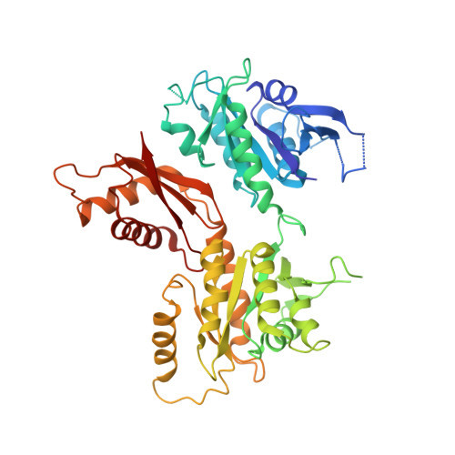 | |
UniProt | |||||
Find proteins for P50743 (Bacillus subtilis (strain 168)) Explore P50743 Go to UniProtKB: P50743 | |||||
Entity Groups | |||||
| Sequence Clusters | 30% Identity50% Identity70% Identity90% Identity95% Identity100% Identity | ||||
| UniProt Group | P50743 | ||||
Sequence AnnotationsExpand | |||||
| |||||
| Ligands 3 Unique | |||||
|---|---|---|---|---|---|
| ID | Chains | Name / Formula / InChI Key | 2D Diagram | 3D Interactions | |
| GNP Query on GNP | C [auth A] | PHOSPHOAMINOPHOSPHONIC ACID-GUANYLATE ESTER C10 H17 N6 O13 P3 UQABYHGXWYXDTK-UUOKFMHZSA-N |  | ||
| PO4 Query on PO4 | B [auth A], E [auth A] | PHOSPHATE ION O4 P NBIIXXVUZAFLBC-UHFFFAOYSA-K |  | ||
| K Query on K | D [auth A] | POTASSIUM ION K NPYPAHLBTDXSSS-UHFFFAOYSA-N |  | ||
| Length ( Å ) | Angle ( ˚ ) |
|---|---|
| a = 50.93 | α = 90 |
| b = 63.88 | β = 97.34 |
| c = 62.45 | γ = 90 |
| Software Name | Purpose |
|---|---|
| REFMAC | refinement |
| XDS | data reduction |
| XSCALE | data scaling |
| PHASER | phasing |
RCSB PDB (citation) is hosted by
RCSB PDB is a member of the

