Structural implications of Ca(2+)-dependent actin-bundling function of human EFhd2/Swiprosin-1.
Park, K.R., Kwon, M.S., An, J.Y., Lee, J.G., Youn, H.S., Lee, Y., Kang, J.Y., Kim, T.G., Lim, J.J., Park, J.S., Lee, S.H., Song, W.K., Cheong, H.K., Jun, C.D., Eom, S.H.(2016) Sci Rep 6: 39095-39095
- PubMed: 27974828
- DOI: https://doi.org/10.1038/srep39095
- Primary Citation of Related Structures:
5I2L, 5I2O, 5I2Q - PubMed Abstract:
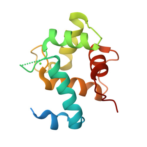
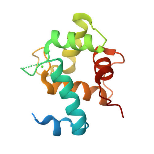
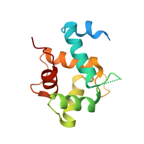
EFhd2/Swiprosin-1 is a cytoskeletal Ca 2+ -binding protein implicated in Ca 2+ -dependent cell spreading and migration in epithelial cells. EFhd2 domain architecture includes an N-terminal disordered region, a PxxP motif, two EF-hands, a ligand mimic helix and a C-terminal coiled-coil domain. We reported previously that EFhd2 displays F-actin bundling activity in the presence of Ca 2+ and this activity depends on the coiled-coil domain and direct interaction of the EFhd2 core region. However, the molecular mechanism for the regulation of F-actin binding and bundling by EFhd2 is unknown. Here, the Ca 2+ -bound crystal structure of the EFhd2 core region is presented and structures of mutants defective for Ca 2+ -binding are also described. These structures and biochemical analyses reveal that the F-actin bundling activity of EFhd2 depends on the structural rigidity of F-actin binding sites conferred by binding of the EF-hands to Ca 2+ . In the absence of Ca 2+ , the EFhd2 core region exhibits local conformational flexibility around the EF-hand domain and C-terminal linker, which retains F-actin binding activity but loses the ability to bundle F-actin. In addition, we establish that dimerisation of EFhd2 via the C-terminal coiled-coil domain, which is necessary for F-actin bundling, occurs through the parallel coiled-coil interaction.
Organizational Affiliation:
School of Life Science, Gwangju Institute of Science and Technology (GIST), 123 Cheomdangwagi-ro, Buk-gu, Gwangju 61005, Republic of Korea.


