5HVF

Crystal Structure of Thrombin-activatable Fibrinolysis Inhibitor in Complex with an Inhibitory Nanobody (VHH-i83)


Experimental Data Snapshot

  • Method: X-RAY DIFFRACTION
  • Resolution: 2.85 Å
  • R-Value Free: 0.230 
  • R-Value Work: 0.192 
  • R-Value Observed: 0.194 

wwPDB Validation   3D Report Full Report


Ligand Structure Quality Assessment 


This is version 2.1 of the entry. See complete history


Literature

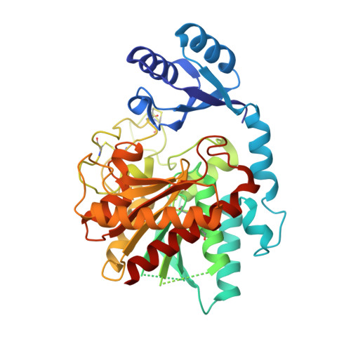
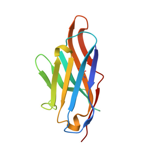
Elucidation of the molecular mechanisms of two nanobodies that inhibit thrombin-activatable fibrinolysis inhibitor activation and activated thrombin-activatable fibrinolysis inhibitor activity.

Zhou, X.Weeks, S.D.Ameloot, P.Callewaert, N.Strelkov, S.V.Declerck, P.J.

(2016) J Thromb Haemost 14: 1629-1638

  • DOI: https://doi.org/10.1111/jth.13381
  • Primary Citation of Related Structures:  
    5HVF, 5HVG, 5HVH

  • PubMed Abstract: 

    Essentials Thrombin-activatable fibrinolysis inhibitor (TAFI) is a risk factor for cardiovascular disorders. TAFI inhibitory nanobodies represent a promising step in developing profibrinolytic therapeutics. We have solved three crystal structures of TAFI in complex with inhibitory nanobodies. Nanobodies inhibit TAFI through distinct mechanisms and represent novel profibrinolytic leads. Background Thrombin-activatable fibrinolysis inhibitor (TAFI) is converted to activated TAFI (TAFIa) by thrombin, plasmin, or the thrombin-thrombomodulin complex (T/TM). TAFIa is antifibrinolytic, and high levels of TAFIa are associated with an increased risk for cardiovascular disorders. TAFI-inhibitory nanobodies represent a promising approach for developing profibrinolytic therapeutics. Objective To elucidate the molecular mechanisms of inhibition of TAFI activation and TAFIa activity by nanobodies with the use of X-ray crystallography and biochemical characterization. Methods and results We selected two nanobodies for cocrystallization with TAFI. VHH-a204 interferes with all TAFI activation modes, whereas VHH-i83 interferes with T/TM-mediated activation and also inhibits TAFIa activity. The 3.05-Å-resolution crystal structure of TAFI-VHH-a204 reveals that the VHH-a204 epitope is localized to the catalytic moiety (CM) in close proximity to the TAFI activation site at Arg92, indicating that VHH-a204 inhibits TAFI activation by steric hindrance. The 2.85-Å-resolution crystal structure of TAFI-VHH-i83 reveals that the VHH-i83 epitope is located close to the presumptive thrombomodulin-binding site in the activation peptide (AP). The structure and supporting biochemical assays suggest that VHH-i83 inhibits TAFIa by bridging the AP to the CM following TAFI activation. In addition, the 3.00-Å-resolution crystal structure of the triple TAFI-VHH-a204-VHH-i83 complex demonstrates that the two nanobodies can simultaneously bind to TAFI. Conclusions This study provides detailed insights into the molecular mechanisms of TAFI inhibition, and reveals a novel mode of TAFIa inhibition. VHH-a204 and VHH-i83 merit further evaluation as potential profibrinolytic therapeutics.


  • Organizational Affiliation

    Department of Pharmaceutical and Pharmacologic Sciences, Laboratory for Therapeutic and Diagnostic Antibodies, KU Leuven, Belgium.


Macromolecules
Find similar proteins by:  (by identity cutoff)  |  3D Structure
Entity ID: 1
MoleculeChains Sequence LengthOrganismDetailsImage
Carboxypeptidase B2401Homo sapiensMutation(s): 4 
Gene Names: CPB2
EC: 3.4.17.20
UniProt & NIH Common Fund Data Resources
Find proteins for Q96IY4 (Homo sapiens)
Explore Q96IY4 
Go to UniProtKB:  Q96IY4
PHAROS:  Q96IY4
GTEx:  ENSG00000080618 
Entity Groups  
Sequence Clusters30% Identity50% Identity70% Identity90% Identity95% Identity100% Identity
UniProt GroupQ96IY4
Sequence Annotations
Expand
  • Reference Sequence
Find similar proteins by:  (by identity cutoff)  |  3D Structure
Entity ID: 2
MoleculeChains Sequence LengthOrganismDetailsImage
VHH-i83119Vicugna pacosMutation(s): 0 
Entity Groups  
Sequence Clusters30% Identity50% Identity70% Identity90% Identity95% Identity100% Identity
Sequence Annotations
Expand
  • Reference Sequence
Oligosaccharides

Help

Entity ID: 3
MoleculeChains Length2D Diagram Glycosylation3D Interactions
alpha-D-mannopyranose-(1-3)-beta-D-mannopyranose-(1-4)-2-acetamido-2-deoxy-beta-D-glucopyranose-(1-4)-2-acetamido-2-deoxy-beta-D-glucopyranose
C
4N-Glycosylation
Glycosylation Resources
GlyTouCan:  G81315DD
GlyCosmos:  G81315DD
GlyGen:  G81315DD
Experimental Data & Validation

Experimental Data

  • Method: X-RAY DIFFRACTION
  • Resolution: 2.85 Å
  • R-Value Free: 0.230 
  • R-Value Work: 0.192 
  • R-Value Observed: 0.194 
  • Space Group: P 21 21 21
Unit Cell:
Length ( Å )Angle ( ˚ )
a = 51.29α = 90
b = 69.978β = 90
c = 165.337γ = 90
Software Package:
Software NamePurpose
BUSTER-TNTrefinement
Aimlessdata scaling
XDSdata reduction
PDB_EXTRACTdata extraction
PHASERphasing

Structure Validation

View Full Validation Report


Ligand Structure Quality Assessment 


Entry History & Funding Information

Deposition Data


Funding OrganizationLocationGrant Number
FWO-VlaanderenBelgiumG072915N

Revision History  (Full details and data files)

  • Version 1.0: 2016-06-22
    Type: Initial release
  • Version 1.1: 2016-08-03
    Changes: Database references
  • Version 1.2: 2016-08-31
    Changes: Database references
  • Version 2.0: 2020-07-29
    Type: Remediation
    Reason: Carbohydrate remediation
    Changes: Atomic model, Data collection, Derived calculations, Structure summary
  • Version 2.1: 2024-01-10
    Changes: Data collection, Database references, Derived calculations, Refinement description, Structure summary