5FM7

Double-heterohexameric rings of full-length Rvb1(ADP)Rvb2(ADP)


Experimental Data Snapshot

  • Method: X-RAY DIFFRACTION
  • Resolution: 2.90 Å
  • R-Value Free: 0.282 
  • R-Value Work: 0.259 
  • R-Value Observed: 0.260 

wwPDB Validation   3D Report Full Report


Ligand Structure Quality Assessment 


This is version 1.2 of the entry. See complete history


Literature

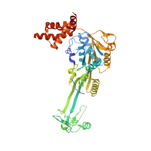
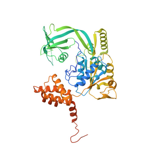
The Combination of X-Ray Crystallography and Cryo-Electron Microscopy Provides Insight Into the Overall Architecture of the Dodecameric Rvb1/Rvb2 Complex.

Silva-Martin, N.Dauden, M.I.Glatt, S.Hoffmann, N.A.Kastritis, P.Bork, P.Beck, M.Muller, C.W.

(2016) PLoS One 11: 46457

  • DOI: https://doi.org/10.1371/journal.pone.0146457
  • Primary Citation of Related Structures:  
    5FM6, 5FM7

  • PubMed Abstract: 

    The Rvb1/Rvb2 complex is an essential component of many cellular pathways. The Rvb1/Rvb2 complex forms a dodecameric assembly where six copies of each subunit form two heterohexameric rings. However, due to conformational variability, the way the two rings pack together is still not fully understood. Here, we present the crystal structure and two cryo-electron microscopy reconstructions of the dodecameric, full-length Rvb1/Rvb2 complex, all showing that the interaction between the two heterohexameric rings is mediated through the Rvb1/Rvb2-specific domain II. Two conformations of the Rvb1/Rvb2 dodecamer are present in solution: a stretched conformation also present in the crystal, and a compact conformation. Novel asymmetric features observed in the reconstruction of the compact conformation provide additional insight into the plasticity of the Rvb1/Rvb2 complex.


  • Organizational Affiliation

    Structural and Computational Biology Unit, European Molecular Biology Laboratory (EMBL), Heidelberg, Germany.


Macromolecules
Find similar proteins by:  (by identity cutoff)  |  3D Structure
Entity ID: 1
MoleculeChains Sequence LengthOrganismDetailsImage
RVB1464Thermochaetoides thermophilaMutation(s): 0 
UniProt
Find proteins for G0RYI5 (Chaetomium thermophilum (strain DSM 1495 / CBS 144.50 / IMI 039719))
Explore G0RYI5 
Go to UniProtKB:  G0RYI5
Entity Groups  
Sequence Clusters30% Identity50% Identity70% Identity90% Identity95% Identity100% Identity
UniProt GroupG0RYI5
Sequence Annotations
Expand
  • Reference Sequence
Find similar proteins by:  (by identity cutoff)  |  3D Structure
Entity ID: 2
MoleculeChains Sequence LengthOrganismDetailsImage
RVB2490Thermochaetoides thermophilaMutation(s): 0 
UniProt
Find proteins for G0RYC2 (Chaetomium thermophilum (strain DSM 1495 / CBS 144.50 / IMI 039719))
Explore G0RYC2 
Go to UniProtKB:  G0RYC2
Entity Groups  
Sequence Clusters30% Identity50% Identity70% Identity90% Identity95% Identity100% Identity
UniProt GroupG0RYC2
Sequence Annotations
Expand
  • Reference Sequence
Small Molecules
Experimental Data & Validation

Experimental Data

  • Method: X-RAY DIFFRACTION
  • Resolution: 2.90 Å
  • R-Value Free: 0.282 
  • R-Value Work: 0.259 
  • R-Value Observed: 0.260 
  • Space Group: H 3 2
Unit Cell:
Length ( Å )Angle ( ˚ )
a = 208.77α = 90
b = 208.77β = 90
c = 138.45γ = 120
Software Package:
Software NamePurpose
PHENIXrefinement
XDSdata reduction
XSCALEdata scaling
PHASERphasing

Structure Validation

View Full Validation Report


Ligand Structure Quality Assessment 


Entry History 

Deposition Data

Revision History  (Full details and data files)

  • Version 1.0: 2016-01-20
    Type: Initial release
  • Version 1.1: 2017-09-27
    Changes: Advisory, Data collection
  • Version 1.2: 2024-01-10
    Changes: Advisory, Data collection, Database references, Derived calculations, Other, Refinement description