5DX5

Crystal structure of methionine gamma-lyase from Clostridium sporogenes


Experimental Data Snapshot

  • Method: X-RAY DIFFRACTION
  • Resolution: 2.37 Å
  • R-Value Free: 0.225 
  • R-Value Work: 0.162 
  • R-Value Observed: 0.165 

wwPDB Validation   3D Report Full Report


Ligand Structure Quality Assessment 


This is version 1.2 of the entry. See complete history


Literature

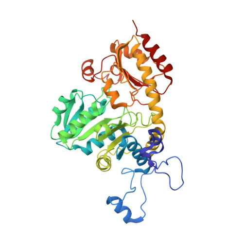
Structure of methionine gamma-lyase from Clostridium sporogenes.

Revtovich, S.Anufrieva, N.Morozova, E.Kulikova, V.Nikulin, A.Demidkina, T.

(2016) Acta Crystallogr F Struct Biol Commun 72: 65-71

  • DOI: https://doi.org/10.1107/S2053230X15023869
  • Primary Citation of Related Structures:  
    5DX5

  • PubMed Abstract: 

    Methionine γ-lyase (MGL) is a pyridoxal 5'-phosphate-dependent enzyme that catalyzes the γ-elimination reaction of L-methionine. The enzyme is a promising target for therapeutic intervention in some anaerobic pathogens and has attracted interest as a potential cancer treatment. The crystal structure of MGL from Clostridium sporogenes has been determined at 2.37 Å resolution. The fold of the protein is similar to those of homologous enzymes from Citrobacter freundii, Entamoeba histolytica, Pseudomonas putida and Trichomonas vaginalis. A comparison of these structures revealed differences in the conformation of two flexible regions of the N- and C-terminal domains involved in the active-site architecture.


  • Organizational Affiliation

    Engelhardt Institute of Molecular Biology, Russian Academy of Sciences, Vavilov str. 32, Moscow 119991, Russian Federation.


Macromolecules
Find similar proteins by:  (by identity cutoff)  |  3D Structure
Entity ID: 1
MoleculeChains Sequence LengthOrganismDetailsImage
Methionine gamma-lyase
A, B
400Clostridium sporogenesMutation(s): 0 
Gene Names: megLCLOSPO_00030
EC: 4.4.1.11
UniProt
Find proteins for J7TA22 (Clostridium sporogenes (strain ATCC 15579))
Explore J7TA22 
Go to UniProtKB:  J7TA22
Entity Groups  
Sequence Clusters30% Identity50% Identity70% Identity90% Identity95% Identity100% Identity
UniProt GroupJ7TA22
Sequence Annotations
Expand
  • Reference Sequence
Experimental Data & Validation

Experimental Data

  • Method: X-RAY DIFFRACTION
  • Resolution: 2.37 Å
  • R-Value Free: 0.225 
  • R-Value Work: 0.162 
  • R-Value Observed: 0.165 
  • Space Group: P 43 21 2
Unit Cell:
Length ( Å )Angle ( ˚ )
a = 91.09α = 90
b = 91.09β = 90
c = 175.929γ = 90
Software Package:
Software NamePurpose
MxCuBEdata collection
XDSdata reduction
SCALAdata scaling
PHASERphasing
PHENIXrefinement

Structure Validation

View Full Validation Report


Ligand Structure Quality Assessment 


Entry History & Funding Information

Deposition Data


Funding OrganizationLocationGrant Number
Russian Scientific FoundationRussian Federation15-14-00009

Revision History  (Full details and data files)

  • Version 1.0: 2016-01-13
    Type: Initial release
  • Version 1.1: 2016-01-20
    Changes: Database references
  • Version 1.2: 2024-01-10
    Changes: Data collection, Database references, Refinement description