5AEE

A bacterial protein structure in glycoside hydrolase family 31


Experimental Data Snapshot

  • Method: X-RAY DIFFRACTION
  • Resolution: 1.85 Å
  • R-Value Free: 0.201 
  • R-Value Work: 0.163 
  • R-Value Observed: 0.164 

wwPDB Validation   3D Report Full Report


Ligand Structure Quality Assessment 


This is version 2.1 of the entry. See complete history


Literature

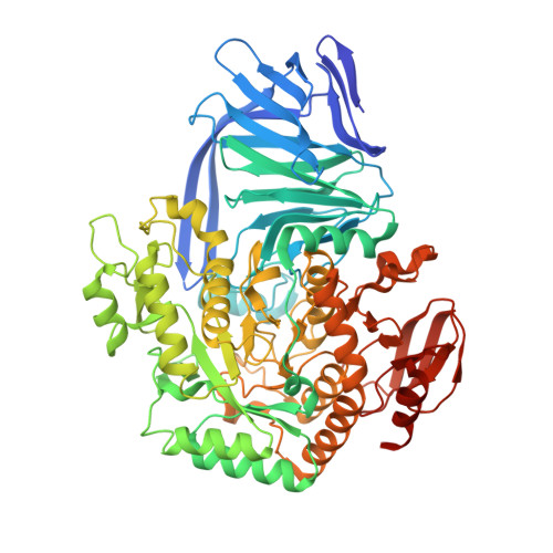
Yihq is a Sulfoquinovosidase that Cleaves Sulfoquinovosyl Diacylglyceride Sulfolipids.

Speciale, G.Jin, Y.Davies, G.J.Williams, S.J.Goddard-Borger, E.D.

(2016) Nat Chem Biol 12: 215

  • DOI: https://doi.org/10.1038/nchembio.2023
  • Primary Citation of Related Structures:  
    5AED, 5AEE, 5AEG

  • PubMed Abstract: 

    Sulfoquinovose is produced by photosynthetic organisms at a rate of 10(10) tons per annum and is degraded by bacteria as a source of carbon and sulfur. We have identified Escherichia coli YihQ as the first dedicated sulfoquinovosidase and the gateway enzyme to sulfoglycolytic pathways. Structural and mutagenesis studies unveiled the sequence signatures for binding the distinguishing sulfonate residue and revealed that sulfoquinovoside degradation is widespread across the tree of life.


  • Organizational Affiliation

    School of Chemistry, University of Melbourne, Parkville, Victoria, Australia.


Macromolecules
Find similar proteins by:  (by identity cutoff)  |  3D Structure
Entity ID: 1
MoleculeChains Sequence LengthOrganismDetailsImage
ALPHA-GLUCOSIDASE YIHQ
A, B
686Escherichia coli K-12Mutation(s): 1 
EC: 3.2.1.20
UniProt
Find proteins for P32138 (Escherichia coli (strain K12))
Explore P32138 
Go to UniProtKB:  P32138
Entity Groups  
Sequence Clusters30% Identity50% Identity70% Identity90% Identity95% Identity100% Identity
UniProt GroupP32138
Sequence Annotations
Expand
  • Reference Sequence
Small Molecules
Ligands 4 Unique
IDChains Name / Formula / InChI Key2D Diagram3D Interactions
NSQ
Query on NSQ

Download Ideal Coordinates CCD File 
E [auth A],
H [auth B]
4-nitrophenyl alpha-D-6-sulfoquinovoside
C12 H15 N O10 S
JRVQBBIDMNDOQV-ZIQFBCGOSA-N
MPD
Query on MPD

Download Ideal Coordinates CCD File 
G [auth B](4S)-2-METHYL-2,4-PENTANEDIOL
C6 H14 O2
SVTBMSDMJJWYQN-YFKPBYRVSA-N
CA
Query on CA

Download Ideal Coordinates CCD File 
C [auth A],
D [auth A],
F [auth B]
CALCIUM ION
Ca
BHPQYMZQTOCNFJ-UHFFFAOYSA-N
CL
Query on CL

Download Ideal Coordinates CCD File 
I [auth B]CHLORIDE ION
Cl
VEXZGXHMUGYJMC-UHFFFAOYSA-M
Experimental Data & Validation

Experimental Data

  • Method: X-RAY DIFFRACTION
  • Resolution: 1.85 Å
  • R-Value Free: 0.201 
  • R-Value Work: 0.163 
  • R-Value Observed: 0.164 
  • Space Group: P 1 21 1
Unit Cell:
Length ( Å )Angle ( ˚ )
a = 77.96α = 90
b = 107.62β = 107.85
c = 103.3γ = 90
Software Package:
Software NamePurpose
REFMACrefinement
REFMACphasing

Structure Validation

View Full Validation Report


Ligand Structure Quality Assessment 


Entry History 

Deposition Data

Revision History  (Full details and data files)

  • Version 1.0: 2016-02-10
    Type: Initial release
  • Version 1.1: 2016-03-02
    Changes: Database references
  • Version 1.2: 2016-03-30
    Changes: Database references
  • Version 1.3: 2019-05-22
    Changes: Data collection, Experimental preparation, Other
  • Version 2.0: 2020-07-29
    Type: Remediation
    Reason: Carbohydrate remediation
    Changes: Atomic model, Derived calculations, Other, Structure summary
  • Version 2.1: 2024-01-10
    Changes: Data collection, Database references, Refinement description, Structure summary