4YPO

Crystal structure of Mycobacterium tuberculosis ketol-acid reductoisomerase in complex with Mg2+


Experimental Data Snapshot

  • Method: X-RAY DIFFRACTION
  • Resolution: 1.00 Å
  • R-Value Free: 0.163 
  • R-Value Work: 0.154 
  • R-Value Observed: 0.154 

wwPDB Validation   3D Report Full Report


This is version 1.3 of the entry. See complete history


Literature

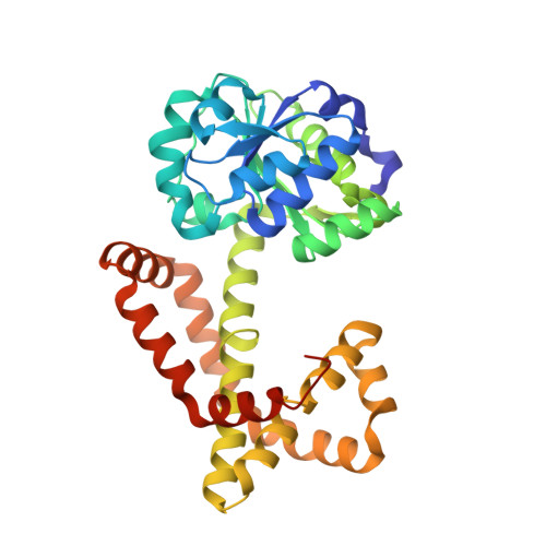
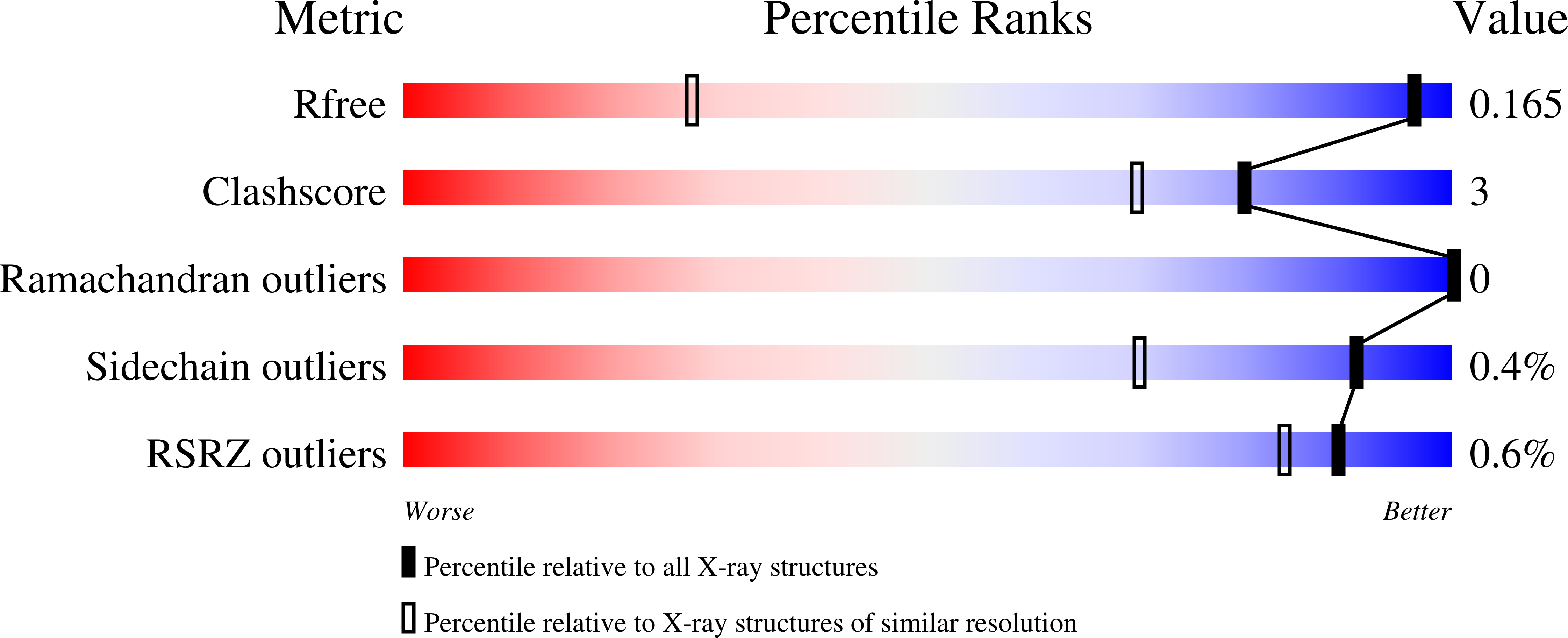
Crystal structure of Mycobacterium tuberculosis ketol-acid reductoisomerase at 1.0 angstrom resolution - a potential target for anti-tuberculosis drug discovery.

Lv, Y.Kandale, A.Wun, S.J.McGeary, R.P.Williams, S.J.Kobe, B.Sieber, V.Schembri, M.A.Schenk, G.Guddat, L.W.

(2016) FEBS J 283: 1184-1196

  • DOI: https://doi.org/10.1111/febs.13672
  • Primary Citation of Related Structures:  
    4YPO

  • PubMed Abstract: 

    The biosynthetic pathway for the branched-chain amino acids is present in plants, fungi and bacteria, but not in animals, making it an attractive target for herbicidal and antimicrobial drug discovery. Ketol-acid reductoisomerase (KARI; EC 1.1.1.86) is the second enzyme in this pathway, converting in a Mg(2+) - and NADPH-dependent reaction either 2-acetolactate or 2-aceto-2-hydroxybutyrate to their corresponding 2,3-dihydroxy-3-alkylbutyrate products. Here, we have determined the crystal structure of Mycobacterium tuberculosis (Mt) KARI, a class I KARI, with two magnesium ions bound in the active site. X-ray data were obtained to 1.0 Å resolution and the final model has an Rfree of 0.163. The structure shows that the active site is solvent-accessible with the two metal ions separated by 4.7 Å. A comparison of this structure with that of Mg(2+) -free Pseudomonas aeruginosa KARI suggests that upon magnesium binding no movement of the N domain relative to the C domain occurs. However, upon formation of the Michaelis complex, as illustrated in the structure of Slackia exigua KARI in complex with NADH.Mg(2+) . N-hydroxy-N-isopropyloxamate (IpOHA, a transition state analog), domain movements and reduction of the metal-metal distance to 3.5 Å are observed. This inherent flexibility therefore appears to be critical for initiation of the KARI-catalyzed reaction. This study provides new insights into the complex structural rearrangements required for activity of KARIs, particularly those belonging to class I, and provides the framework for the rational design of Mt KARI inhibitors that can be tested as novel antituberculosis agents.


  • Organizational Affiliation

    School of Chemistry and Molecular Biosciences and Australian Infectious Disease Research Centre, University of Queensland, Brisbane, Australia.


Macromolecules
Find similar proteins by:  (by identity cutoff)  |  3D Structure
Entity ID: 1
MoleculeChains Sequence LengthOrganismDetailsImage
Ketol-acid reductoisomerase
A, B
333Mycobacterium tuberculosisMutation(s): 0 
Gene Names: ilvCIQ45_13380IU12_14180IU13_13535IU21_13700T209_13330
EC: 1.1.1.86
UniProt
Find proteins for P9WKJ7 (Mycobacterium tuberculosis (strain ATCC 25618 / H37Rv))
Explore P9WKJ7 
Go to UniProtKB:  P9WKJ7
Entity Groups  
Sequence Clusters30% Identity50% Identity70% Identity90% Identity95% Identity100% Identity
UniProt GroupP9WKJ7
Sequence Annotations
Expand
  • Reference Sequence
Small Molecules
Ligands 3 Unique
IDChains Name / Formula / InChI Key2D Diagram3D Interactions
CL
Query on CL

Download Ideal Coordinates CCD File 
G [auth A],
K [auth A]
CHLORIDE ION
Cl
VEXZGXHMUGYJMC-UHFFFAOYSA-M
MG
Query on MG

Download Ideal Coordinates CCD File 
C [auth A]
D [auth A]
E [auth A]
F [auth A]
H [auth A]
C [auth A],
D [auth A],
E [auth A],
F [auth A],
H [auth A],
I [auth A],
J [auth A],
L [auth A],
M [auth B],
N [auth B],
O [auth B],
P [auth B],
Q [auth B]
MAGNESIUM ION
Mg
JLVVSXFLKOJNIY-UHFFFAOYSA-N
NA
Query on NA

Download Ideal Coordinates CCD File 
R [auth B]SODIUM ION
Na
FKNQFGJONOIPTF-UHFFFAOYSA-N
Experimental Data & Validation

Experimental Data

  • Method: X-RAY DIFFRACTION
  • Resolution: 1.00 Å
  • R-Value Free: 0.163 
  • R-Value Work: 0.154 
  • R-Value Observed: 0.154 
  • Space Group: C 1 2 1
Unit Cell:
Length ( Å )Angle ( ˚ )
a = 157.697α = 90
b = 54.797β = 123.25
c = 92.23γ = 90
Software Package:
Software NamePurpose
PHENIXrefinement
HKL-2000data reduction
PHASERphasing

Structure Validation

View Full Validation Report


Entry History 

Deposition Data

Revision History  (Full details and data files)

  • Version 1.0: 2016-02-17
    Type: Initial release
  • Version 1.1: 2016-03-02
    Changes: Database references
  • Version 1.2: 2016-04-27
    Changes: Database references
  • Version 1.3: 2024-02-28
    Changes: Data collection, Database references, Derived calculations