4Y66

Crystal structure of Giardia lamblia Hop2-Mnd1 complex


Experimental Data Snapshot

  • Method: X-RAY DIFFRACTION
  • Resolution: 3.20 Å
  • R-Value Free: 0.286 
  • R-Value Work: 0.239 
  • R-Value Observed: 0.241 

wwPDB Validation   3D Report Full Report


This is version 1.2 of the entry. See complete history


Literature

Crystal structure of Hop2-Mnd1 and mechanistic insights into its role in meiotic recombination

Kang, H.A.Shin, H.C.Kalantzi, A.S.Toseland, C.P.Kim, H.M.Gruber, S.Peraro, M.D.Oh, B.H.

(2015) Nucleic Acids Res 43: 3841-3856

  • DOI: https://doi.org/10.1093/nar/gkv172
  • Primary Citation of Related Structures:  
    4Y66

  • PubMed Abstract: 

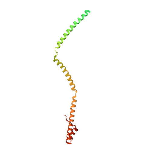
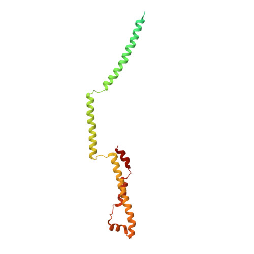
    In meiotic DNA recombination, the Hop2-Mnd1 complex promotes Dmc1-mediated single-stranded DNA (ssDNA) invasion into homologous chromosomes to form a synaptic complex by a yet-unclear mechanism. Here, the crystal structure of Hop2-Mnd1 reveals that it forms a curved rod-like structure consisting of three leucine zippers and two kinked junctions. One end of the rod is linked to two juxtaposed winged-helix domains, and the other end is capped by extra α-helices to form a helical bundle-like structure. Deletion analysis shows that the helical bundle-like structure is sufficient for interacting with the Dmc1-ssDNA nucleofilament, and molecular modeling suggests that the curved rod could be accommodated into the helical groove of the nucleofilament. Remarkably, the winged-helix domains are juxtaposed at fixed relative orientation, and their binding to DNA is likely to perturb the base pairing according to molecular simulations. These findings allow us to propose a model explaining how Hop2-Mnd1 juxtaposes Dmc1-bound ssDNA with distorted recipient double-stranded DNA and thus facilitates strand invasion.


  • Organizational Affiliation

    Department of Biological Sciences, KAIST Institute for the Biocentury, Cancer Metastasis Control Center, Korea Advanced Institute of Science and Technology, Daejeon 305-701, Korea.


Macromolecules
Find similar proteins by:  (by identity cutoff)  |  3D Structure
Entity ID: 1
MoleculeChains Sequence LengthOrganismDetailsImage
Mnd1
A, C, E
203Giardia lamblia ATCC 50803Mutation(s): 0 
Gene Names: GL50803_6626
UniProt
Find proteins for E2RTU1 (Giardia intestinalis (strain ATCC 50803 / WB clone C6))
Explore E2RTU1 
Go to UniProtKB:  E2RTU1
Entity Groups  
Sequence Clusters30% Identity50% Identity70% Identity90% Identity95% Identity100% Identity
UniProt GroupE2RTU1
Sequence Annotations
Expand
  • Reference Sequence
Find similar proteins by:  (by identity cutoff)  |  3D Structure
Entity ID: 2
MoleculeChains Sequence LengthOrganismDetailsImage
Putative tbpip family protein
B, D, F
231Giardia intestinalisMutation(s): 0 
Gene Names: GSB_17044
UniProt
Find proteins for V6TR15 (Giardia intestinalis)
Explore V6TR15 
Go to UniProtKB:  V6TR15
Entity Groups  
Sequence Clusters30% Identity50% Identity70% Identity90% Identity95% Identity100% Identity
UniProt GroupV6TR15
Sequence Annotations
Expand
  • Reference Sequence
Experimental Data & Validation

Experimental Data

  • Method: X-RAY DIFFRACTION
  • Resolution: 3.20 Å
  • R-Value Free: 0.286 
  • R-Value Work: 0.239 
  • R-Value Observed: 0.241 
  • Space Group: C 1 2 1
Unit Cell:
Length ( Å )Angle ( ˚ )
a = 117.847α = 90
b = 69.066β = 95.34
c = 292.137γ = 90
Software Package:
Software NamePurpose
PHENIXrefinement
HKL-2000data reduction
HKL-2000data scaling
PHENIXphasing

Structure Validation

View Full Validation Report


Entry History 

Deposition Data

Revision History  (Full details and data files)

  • Version 1.0: 2015-03-18
    Type: Initial release
  • Version 1.1: 2015-05-06
    Changes: Database references
  • Version 1.2: 2024-03-20
    Changes: Data collection, Database references, Derived calculations, Source and taxonomy