Molecular Details of Olfactomedin Domains Provide Pathway to Structure-Function Studies.
Hill, S.E., Donegan, R.K., Nguyen, E., Desai, T.M., Lieberman, R.L.(2015) PLoS One 10: e0130888-e0130888
- PubMed: 26121352
- DOI: https://doi.org/10.1371/journal.pone.0130888
- Primary Citation of Related Structures:
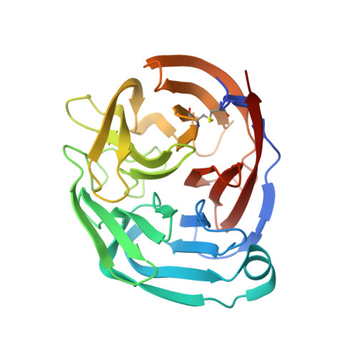
4XAT, 4XAV - PubMed Abstract:
Olfactomedin (OLF) domains are found within extracellular, multidomain proteins in numerous tissues of multicellular organisms. Even though these proteins have been implicated in human disorders ranging from cancers to attention deficit disorder to glaucoma, little is known about their structure(s) and function(s). Here we biophysically, biochemically, and structurally characterize OLF domains from H. sapiens olfactomedin-1 (npoh-OLF, also called noelin, pancortin, OLFM1, and hOlfA), and M. musculus gliomedin (glio-OLF, also called collomin, collmin, and CRG-L2), and compare them with available structures of myocilin (myoc-OLF) recently reported by us and R. norvegicus glio-OLF and M. musculus latrophilin-3 (lat3-OLF) by others. Although the five-bladed β-propeller architecture remains unchanged, numerous physicochemical characteristics differ among these OLF domains. First, npoh-OLF and glio-OLF exhibit prominent, yet distinct, positive surface charges and copurify with polynucleotides. Second, whereas npoh-OLF and myoc-OLF exhibit thermal stabilities typical of human proteins near 55°C, and most myoc-OLF variants are destabilized and highly prone to aggregation, glio-OLF is nearly 20°C more stable and significantly more resistant to chemical denaturation. Phylogenetically, glio-OLF is most similar to primitive OLFs, and structurally, glio-OLF is missing distinguishing features seen in OLFs such as the disulfide bond formed by N- and C- terminal cysteines, the sequestered Ca2+ ion within the propeller central hydrophilic cavity, and a key loop-stabilizing cation-π interaction on the top face of npoh-OLF and myoc-OLF. While deciphering the explicit biological functions, ligands, and binding partners for OLF domains will likely continue to be a challenging long-term experimental pursuit, we used structural insights gained here to generate a new antibody selective for myoc-OLF over npoh-OLF and glio-OLF as a first step in overcoming the impasse in detailed functional characterization of these biomedically important protein domains.
Organizational Affiliation:
School of Chemistry & Biochemistry, Georgia Institute of Technology, Atlanta, Georgia, United States of America.