4URR

Tailspike protein of Sf6 bacteriophage bound to Shigella flexneri O- antigen octasaccharide fragment


Experimental Data Snapshot

  • Method: X-RAY DIFFRACTION
  • Resolution: 1.95 Å
  • R-Value Free: 0.236 
  • R-Value Work: 0.196 
  • R-Value Observed: 0.198 

wwPDB Validation   3D Report Full Report


This is version 2.1 of the entry. See complete history


Literature

Bacteriophage Tailspikes and Bacterial O-Antigens as a Model System to Study Weak-Affinity Protein-Polysaccharide Interactions.

Kang, Y.Gohlke, U.Engstrom, O.Hamark, C.Scheidt, T.Kunstmann, S.Heinemann, U.Widmalm, G.Santer, M.Barbirz, S.

(2016) J Am Chem Soc 138: 9109-9118

  • DOI: https://doi.org/10.1021/jacs.6b00240
  • Primary Citation of Related Structures:  
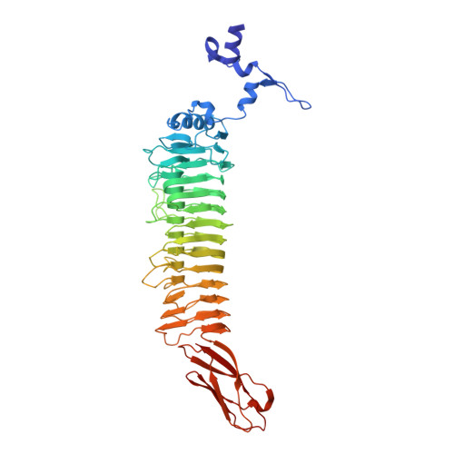
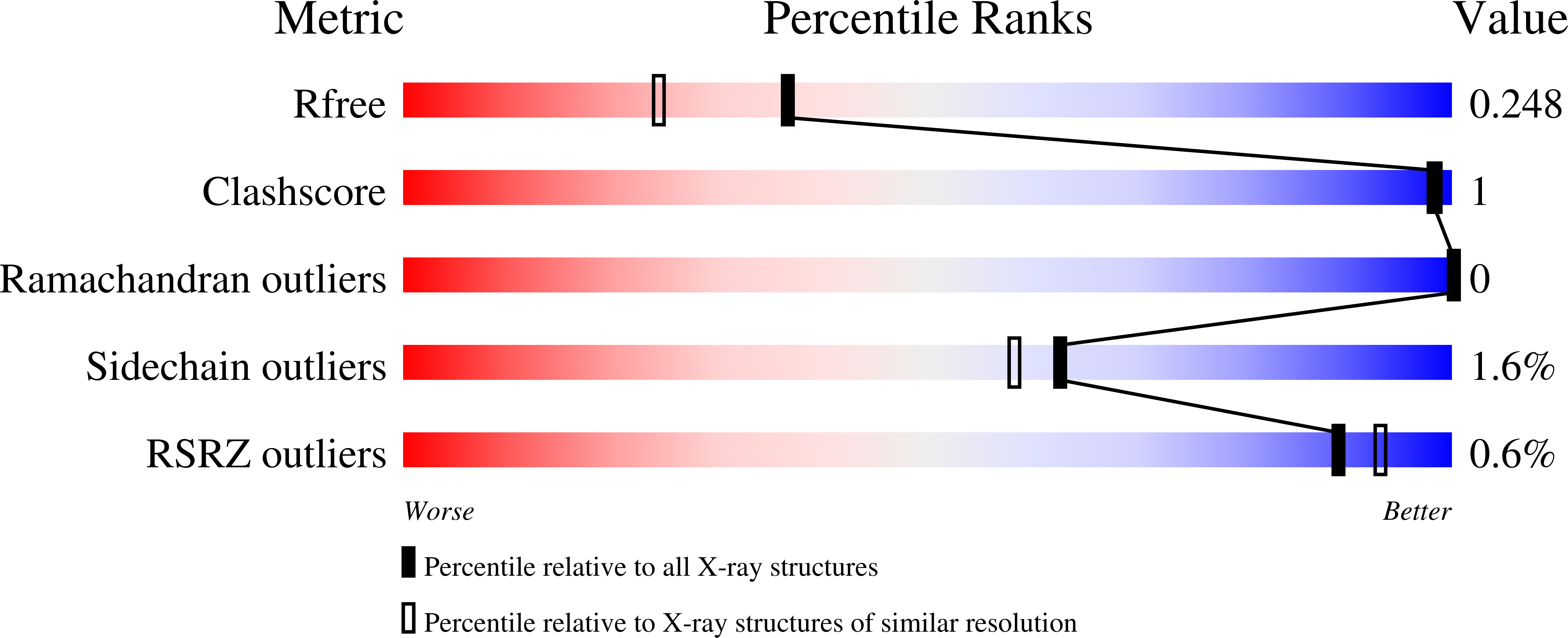
    4URR

  • PubMed Abstract: 

    Understanding interactions of bacterial surface polysaccharides with receptor protein scaffolds is important for the development of antibiotic therapies. The corresponding protein recognition domains frequently form low-affinity complexes with polysaccharides that are difficult to address with experimental techniques due to the conformational flexibility of the polysaccharide. In this work, we studied the tailspike protein (TSP) of the bacteriophage Sf6. Sf6TSP binds and hydrolyzes the high-rhamnose, serotype Y O-antigen polysaccharide of the Gram-negative bacterium Shigella flexneri (S. flexneri) as a first step of bacteriophage infection. Spectroscopic analyses and enzymatic cleavage assays confirmed that Sf6TSP binds long stretches of this polysaccharide. Crystal structure analysis and saturation transfer difference (STD) NMR spectroscopy using an enhanced method to interpret the data permitted the detailed description of affinity contributions and flexibility in an Sf6TSP-octasaccharide complex. Dodecasaccharide fragments corresponding to three repeating units of the O-antigen in complex with Sf6TSP were studied computationally by molecular dynamics simulations. They showed that distortion away from the low-energy solution conformation found in the octasaccharide complex is necessary for ligand binding. This is in agreement with a weak-affinity functional polysaccharide-protein contact that facilitates correct placement and thus hydrolysis of the polysaccharide close to the catalytic residues. Our simulations stress that the flexibility of glycan epitopes together with a small number of specific protein contacts provide the driving force for Sf6TSP-polysaccharide complex formation in an overall weak-affinity interaction system.


  • Organizational Affiliation

    Max Planck Institute of Colloids and Interfaces , Am Mühlenberg 1, 14476 Potsdam, Germany.


Macromolecules
Find similar proteins by:  (by identity cutoff)  |  3D Structure
Entity ID: 1
MoleculeChains Sequence LengthOrganismDetailsImage
BIFUNCTIONAL TAIL PROTEIN
A, B, C, D, E
A, B, C, D, E, F
514Lederbergvirus Sf6Mutation(s): 2 
EC: 3.2.1
UniProt
Find proteins for Q9XJP3 (Shigella phage Sf6)
Explore Q9XJP3 
Go to UniProtKB:  Q9XJP3
Entity Groups  
Sequence Clusters30% Identity50% Identity70% Identity90% Identity95% Identity100% Identity
UniProt GroupQ9XJP3
Sequence Annotations
Expand
  • Reference Sequence
Oligosaccharides

Help

Entity ID: 2
MoleculeChains Length2D Diagram Glycosylation3D Interactions
alpha-L-rhamnopyranose-(1-3)-2-acetamido-2-deoxy-beta-D-glucopyranose-(1-2)-alpha-L-rhamnopyranose-(1-2)-alpha-L-rhamnopyranose-(1-3)-alpha-L-rhamnopyranose-(1-3)-2-acetamido-2-deoxy-beta-D-glucopyranose-(1-2)-alpha-L-rhamnopyranose-(1-2)-alpha-L-rhamnopyranose
G, H, I, J, K
G, H, I, J, K, L
8N/A
Glycosylation Resources
GlyTouCan:  G27179CQ
GlyCosmos:  G27179CQ
Small Molecules
Ligands 3 Unique
IDChains Name / Formula / InChI Key2D Diagram3D Interactions
PO4
Query on PO4

Download Ideal Coordinates CCD File 
BA [auth F],
DA [auth F],
P [auth A],
Q [auth B]
PHOSPHATE ION
O4 P
NBIIXXVUZAFLBC-UHFFFAOYSA-K
EDO
Query on EDO

Download Ideal Coordinates CCD File 
AA [auth E],
EA [auth F],
S [auth B],
T [auth B],
Z [auth E]
1,2-ETHANEDIOL
C2 H6 O2
LYCAIKOWRPUZTN-UHFFFAOYSA-N
MN
Query on MN

Download Ideal Coordinates CCD File 
CA [auth F]
M [auth A]
N [auth A]
O [auth A]
R [auth B]
CA [auth F],
M [auth A],
N [auth A],
O [auth A],
R [auth B],
U [auth C],
V [auth D],
W [auth D],
X [auth D],
Y [auth E]
MANGANESE (II) ION
Mn
WAEMQWOKJMHJLA-UHFFFAOYSA-N
Experimental Data & Validation

Experimental Data

  • Method: X-RAY DIFFRACTION
  • Resolution: 1.95 Å
  • R-Value Free: 0.236 
  • R-Value Work: 0.196 
  • R-Value Observed: 0.198 
  • Space Group: P 1 21 1
Unit Cell:
Length ( Å )Angle ( ˚ )
a = 82.987α = 90
b = 138.895β = 92.05
c = 136.073γ = 90
Software Package:
Software NamePurpose
BUSTERrefinement
XDSdata reduction
SCALAdata scaling
PHASERphasing

Structure Validation

View Full Validation Report


Entry History 

Deposition Data

Revision History  (Full details and data files)

  • Version 1.0: 2015-07-15
    Type: Initial release
  • Version 1.1: 2020-03-11
    Changes: Data collection, Database references, Derived calculations, Other
  • Version 2.0: 2020-07-29
    Type: Remediation
    Reason: Carbohydrate remediation
    Changes: Advisory, Atomic model, Data collection, Derived calculations, Structure summary
  • Version 2.1: 2024-01-10
    Changes: Data collection, Database references, Refinement description, Structure summary