4QTR

Computational design of co-assembling protein-DNA nanowires


Experimental Data Snapshot

  • Method: X-RAY DIFFRACTION
  • Resolution: 3.20 Å
  • R-Value Free: 0.322 
  • R-Value Work: 0.264 
  • R-Value Observed: 0.270 

wwPDB Validation   3D Report Full Report


This is version 1.3 of the entry. See complete history


Literature

Computational design of co-assembling protein-DNA nanowires.

Mou, Y.Yu, J.Y.Wannier, T.M.Guo, C.L.Mayo, S.L.

(2015) Nature 525: 230-233

  • DOI: https://doi.org/10.1038/nature14874
  • Primary Citation of Related Structures:  
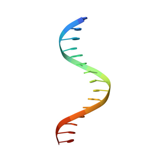
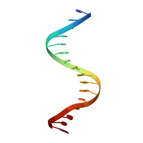
    4QTR

  • PubMed Abstract: 

    Biomolecular self-assemblies are of great interest to nanotechnologists because of their functional versatility and their biocompatibility. Over the past decade, sophisticated single-component nanostructures composed exclusively of nucleic acids, peptides and proteins have been reported, and these nanostructures have been used in a wide range of applications, from drug delivery to molecular computing. Despite these successes, the development of hybrid co-assemblies of nucleic acids and proteins has remained elusive. Here we use computational protein design to create a protein-DNA co-assembling nanomaterial whose assembly is driven via non-covalent interactions. To achieve this, a homodimerization interface is engineered onto the Drosophila Engrailed homeodomain (ENH), allowing the dimerized protein complex to bind to two double-stranded DNA (dsDNA) molecules. By varying the arrangement of protein-binding sites on the dsDNA, an irregular bulk nanoparticle or a nanowire with single-molecule width can be spontaneously formed by mixing the protein and dsDNA building blocks. We characterize the protein-DNA nanowire using fluorescence microscopy, atomic force microscopy and X-ray crystallography, confirming that the nanowire is formed via the proposed mechanism. This work lays the foundation for the development of new classes of protein-DNA hybrid materials. Further applications can be explored by incorporating DNA origami, DNA aptamers and/or peptide epitopes into the protein-DNA framework presented here.


  • Organizational Affiliation

    Division of Chemistry and Chemical Engineering, California Institute of Technology, Pasadena, California 91125, USA.


Macromolecules

Find similar proteins by:  (by identity cutoff)  |  3D Structure
Entity ID: 1
MoleculeChains Sequence LengthOrganismDetailsImage
dualENH
A, B, C, D
72Drosophila melanogasterMutation(s): 0 
Entity Groups  
Sequence Clusters30% Identity50% Identity70% Identity90% Identity95% Identity100% Identity
Sequence Annotations
Expand
  • Reference Sequence

Find similar nucleic acids by:  Sequence   |   3D Structure  

Entity ID: 2
MoleculeChains LengthOrganismImage
DNA (5'-D(P*GP*TP*GP*TP*AP*AP*TP*TP*TP*AP*AP*TP*TP*TP*CP*C)-3')E,
F [auth G]
16N/A
Sequence Annotations
Expand
  • Reference Sequence

Find similar nucleic acids by:  Sequence   |   3D Structure  

Entity ID: 3
MoleculeChains LengthOrganismImage
DNA (5'-D(P*CP*GP*GP*AP*AP*AP*TP*TP*AP*AP*AP*TP*TP*AP*CP*A)-3')G [auth F],
H
16N/A
Sequence Annotations
Expand
  • Reference Sequence
Experimental Data & Validation

Experimental Data

  • Method: X-RAY DIFFRACTION
  • Resolution: 3.20 Å
  • R-Value Free: 0.322 
  • R-Value Work: 0.264 
  • R-Value Observed: 0.270 
  • Space Group: P 42 2 2
Unit Cell:
Length ( Å )Angle ( ˚ )
a = 90.098α = 90
b = 90.098β = 90
c = 158.923γ = 90
Software Package:
Software NamePurpose
CrystalCleardata collection
PHASESphasing
PHENIXrefinement
DENZOdata reduction
SCALAdata scaling

Structure Validation

View Full Validation Report


Entry History 

Deposition Data

Revision History  (Full details and data files)

  • Version 1.0: 2015-07-29
    Type: Initial release
  • Version 1.1: 2015-09-09
    Changes: Database references
  • Version 1.2: 2015-09-30
    Changes: Database references
  • Version 1.3: 2023-09-20
    Changes: Data collection, Database references, Refinement description