4QGN

Human acireductone dioxygenase with iron ion and L-methionine in active center


Experimental Data Snapshot

  • Method: X-RAY DIFFRACTION
  • Resolution: 3.05 Å
  • R-Value Free: 0.228 
  • R-Value Work: 0.178 
  • R-Value Observed: 0.180 

wwPDB Validation   3D Report Full Report


Ligand Structure Quality Assessment 


This is version 1.5 of the entry. See complete history


Literature

On the Structure and Reaction Mechanism of Human Acireductone Dioxygenase.

Milaczewska, A.Kot, E.Amaya, J.A.Makris, T.M.Zajac, M.Korecki, J.Chumakov, A.Trzewik, B.Kedracka-Krok, S.Minor, W.Chruszcz, M.Borowski, T.

(2018) Chemistry 24: 5225-5237

  • DOI: https://doi.org/10.1002/chem.201704617
  • Primary Citation of Related Structures:  
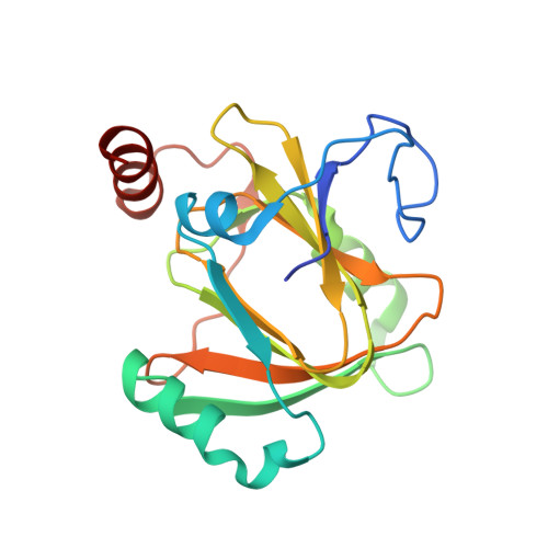
    4QGN

  • PubMed Abstract: 

    Acireductone dioxygenase (ARD) is an intriguing enzyme from the methionine salvage pathway that is capable of catalysing two different oxidation reactions with the same substrate depending on the type of the metal ion in the active site. To date, the structural information regarding the ARD-acireductone complex is limited and possible reaction mechanisms are still under debate. The results of joint experimental and computational studies undertaken to advance knowledge about ARD are reported. The crystal structure of an ARD from Homo sapiens was determined with selenomethionine. EPR spectroscopy suggested that binding acireductone triggers one protein residue to dissociate from Fe 2+ , which allows NO (and presumably O 2 ) to bind directly to the metal. Mössbauer spectroscopic data (interpreted with the aid of DFT calculations) was consistent with bidentate binding of acireductone to Fe 2+ and concomitant dissociation of His88 from the metal. Major features of Fe vibrational spectra obtained for the native enzyme and upon addition of acireductone were reproduced by QM/MM calculations for the proposed models. A computational (QM/MM) study of the reaction mechanisms suggests that Fe 2+ promotes O-O bond homolysis, which elicits cleavage of the C1-C2 bond of the substrate. Higher M 3+ /M 2+ redox potentials of other divalent metals do not support this pathway, and instead the reaction proceeds similarly to the key reaction step in the quercetin 2,3-dioxygenase mechanism.


  • Organizational Affiliation

    Jerzy Haber Institute of Catalysis and Surface Chemistry, Polish Academy of Sciences, Niezapominajek 8, 30-239, Krakow, Poland.


Macromolecules
Find similar proteins by:  (by identity cutoff)  |  3D Structure
Entity ID: 1
MoleculeChains Sequence LengthOrganismDetailsImage
1,2-dihydroxy-3-keto-5-methylthiopentene dioxygenase183Homo sapiensMutation(s): 0 
Gene Names: ADI1HMFT1638MTCBP1NP_060739
EC: 1.13.11.54
UniProt & NIH Common Fund Data Resources
Find proteins for Q9BV57 (Homo sapiens)
Explore Q9BV57 
Go to UniProtKB:  Q9BV57
PHAROS:  Q9BV57
GTEx:  ENSG00000182551 
Entity Groups  
Sequence Clusters30% Identity50% Identity70% Identity90% Identity95% Identity100% Identity
UniProt GroupQ9BV57
Sequence Annotations
Expand
  • Reference Sequence
Small Molecules
Ligands 5 Unique
IDChains Name / Formula / InChI Key2D Diagram3D Interactions
MSE
Query on MSE

Download Ideal Coordinates CCD File 
E [auth A]SELENOMETHIONINE
C5 H11 N O2 Se
RJFAYQIBOAGBLC-BYPYZUCNSA-N
SO4
Query on SO4

Download Ideal Coordinates CCD File 
F [auth A],
G [auth A],
H [auth A],
I [auth A]
SULFATE ION
O4 S
QAOWNCQODCNURD-UHFFFAOYSA-L
ACT
Query on ACT

Download Ideal Coordinates CCD File 
J [auth A]ACETATE ION
C2 H3 O2
QTBSBXVTEAMEQO-UHFFFAOYSA-M
FE
Query on FE

Download Ideal Coordinates CCD File 
B [auth A]FE (III) ION
Fe
VTLYFUHAOXGGBS-UHFFFAOYSA-N
CL
Query on CL

Download Ideal Coordinates CCD File 
C [auth A],
D [auth A]
CHLORIDE ION
Cl
VEXZGXHMUGYJMC-UHFFFAOYSA-M
Experimental Data & Validation

Experimental Data

  • Method: X-RAY DIFFRACTION
  • Resolution: 3.05 Å
  • R-Value Free: 0.228 
  • R-Value Work: 0.178 
  • R-Value Observed: 0.180 
  • Space Group: P 64
Unit Cell:
Length ( Å )Angle ( ˚ )
a = 105.195α = 90
b = 105.195β = 90
c = 40.822γ = 120
Software Package:
Software NamePurpose
HKL-3000data collection
DENZOdata reduction
SCALEPACKdata scaling
REFMACrefinement
Cootmodel building
HKL-3000phasing
CCP4refinement
HKL-3000data reduction
HKL-3000data scaling

Structure Validation

View Full Validation Report


Ligand Structure Quality Assessment 


Entry History 

Deposition Data

Revision History  (Full details and data files)

  • Version 1.0: 2015-05-27
    Type: Initial release
  • Version 1.1: 2017-11-22
    Changes: Refinement description
  • Version 1.2: 2022-04-13
    Changes: Database references, Derived calculations, Structure summary
  • Version 1.3: 2023-08-09
    Changes: Data collection, Database references
  • Version 1.4: 2023-09-20
    Changes: Refinement description
  • Version 1.5: 2023-12-06
    Changes: Data collection