4N6S

Crystals of cross-linked stabilized and functional Phycobilisomes: only phycocyanin rods contribute to diffraction.


Experimental Data Snapshot

  • Method: X-RAY DIFFRACTION
  • Resolution: 2.40 Å
  • R-Value Free: 0.235 
  • R-Value Work: 0.192 

wwPDB Validation   3D Report Full Report


Ligand Structure Quality Assessment 


This is version 1.2 of the entry. See complete history


Literature

Structural studies show energy transfer within stabilized phycobilisomes independent of the mode of rod-core assembly.

David, L.Prado, M.Arteni, A.A.Elmlund, D.A.Blankenship, R.E.Adir, N.

(2014) Biochim Biophys Acta 1837: 385-395

  • DOI: https://doi.org/10.1016/j.bbabio.2013.12.014
  • Primary Citation of Related Structures:  
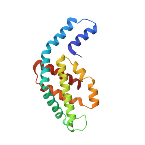
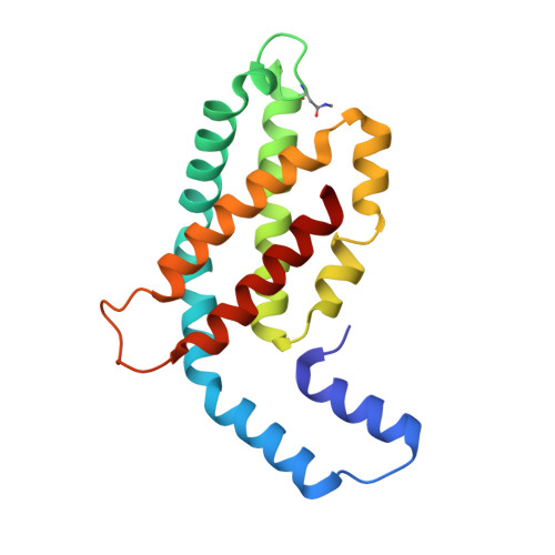
    4N6S

  • PubMed Abstract: 

    The major light harvesting complex in cyanobacteria and red algae is the phycobilisome (PBS), comprised of hundreds of seemingly similar chromophores, which are protein bound and assembled in a fashion that enables highly efficient uni-directional energy transfer to reaction centers. The PBS is comprised of a core containing 2-5 cylinders surrounded by 6-8 rods, and a number of models have been proposed describing the PBS structure. One of the most critical steps in the functionality of the PBS is energy transfer from the rod substructures to the core substructure. In this study we compare the structural and functional characteristics of high-phosphate stabilized PBS (the standard fashion of stabilization of isolated complexes) with cross-linked PBS in low ionic strength buffer from two cyanobacterial species, Thermosynechococcus vulcanus and Acaryochloris marina. We show that chemical cross-linking preserves efficient energy transfer from the phycocyanin containing rods to the allophycocyanin containing cores with fluorescent emission from the terminal emitters. However, this energy transfer is shown to exist in PBS complexes of different structures as characterized by determination of a 2.4Å structure by X-ray crystallography, single crystal confocal microscopy, mass spectrometry and transmission electron microscopy of negatively stained and cryogenically preserved complexes. We conclude that the PBS has intrinsic structural properties that enable efficient energy transfer from rod substructures to the core substructures without requiring a single unique structure. We discuss the significance of our observations on the functionality of the PBS in vivo.


  • Organizational Affiliation

    Schulich Faculty of Chemistry, Technion-Israel Institute of Technology, Haifa 32000, Israel.


Macromolecules
Find similar proteins by:  (by identity cutoff)  |  3D Structure
Entity ID: 1
MoleculeChains Sequence LengthOrganismDetailsImage
C-phycocyanin alpha subunit162Thermostichus vulcanusMutation(s): 0 
UniProt
Find proteins for Q9AM02 (Thermostichus vulcanus)
Explore Q9AM02 
Go to UniProtKB:  Q9AM02
Entity Groups  
Sequence Clusters30% Identity50% Identity70% Identity90% Identity95% Identity100% Identity
UniProt GroupQ9AM02
Sequence Annotations
Expand
  • Reference Sequence
Find similar proteins by:  (by identity cutoff)  |  3D Structure
Entity ID: 2
MoleculeChains Sequence LengthOrganismDetailsImage
C-phycocyanin beta subunit172Thermostichus vulcanusMutation(s): 0 
UniProt
Find proteins for Q71RW8 (Thermostichus vulcanus)
Explore Q71RW8 
Go to UniProtKB:  Q71RW8
Entity Groups  
Sequence Clusters30% Identity50% Identity70% Identity90% Identity95% Identity100% Identity
UniProt GroupQ71RW8
Sequence Annotations
Expand
  • Reference Sequence
Small Molecules
Modified Residues  1 Unique
IDChains TypeFormula2D DiagramParent
MEN
Query on MEN
B
L-PEPTIDE LINKINGC5 H10 N2 O3ASN
Experimental Data & Validation

Experimental Data

  • Method: X-RAY DIFFRACTION
  • Resolution: 2.40 Å
  • R-Value Free: 0.235 
  • R-Value Work: 0.192 
  • Space Group: H 3 2
Unit Cell:
Length ( Å )Angle ( ˚ )
a = 188.07α = 90
b = 188.07β = 90
c = 60.38γ = 120
Software Package:
Software NamePurpose
DNAdata collection
PHASERphasing
CNSrefinement
MOSFLMdata reduction
SCALAdata scaling

Structure Validation

View Full Validation Report


Ligand Structure Quality Assessment 


Entry History 

Deposition Data

Revision History  (Full details and data files)

  • Version 1.0: 2014-01-22
    Type: Initial release
  • Version 1.1: 2014-02-12
    Changes: Database references
  • Version 1.2: 2023-09-20
    Changes: Data collection, Database references, Derived calculations, Refinement description