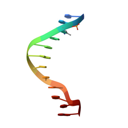
Comparison of the structural and dynamic effects of 5-methylcytosine and 5-chlorocytosine in a CpG dinucleotide sequence.
Theruvathu, J.A., Yin, Y.W., Pettitt, B.M., Sowers, L.C.(2013) Biochemistry 52: 8590-8598
- PubMed: 24147911
- DOI: https://doi.org/10.1021/bi400980c
- Primary Citation of Related Structures:
4MGW, 4MKW - PubMed Abstract:
Inflammation-mediated reactive molecules can result in an array of oxidized and halogenated DNA-damage products, including 5-chlorocytosine ((Cl)C). Previous studies have shown that (Cl)C can mimic 5-methylcytosine ((m)C) and act as a fraudulent epigenetic signal, promoting the methylation of previously unmethylated DNA sequences. Although the 5-halouracils are good substrates for base-excision repair, no repair activity has yet been identified for (Cl)C. Because of the apparent biochemical similarities of (m)C and (Cl)C, we have investigated the effects of (m)C and (Cl)C substitution on oligonucleotide structure and dynamics. In this study, we have constructed oligonucleotide duplexes containing C, (Cl)C, and (m)C within a CpG dinucleotide. The thermal and thermodynamic stability of these duplexes were found to be experimentally indistinguishable. Crystallographic structures of duplex oligonucleotides containing (m)C or (Cl)C were determined to 1.2 and 1.9 Å resolution, respectively. Both duplexes are B-form and are superimposable on a previously determined structure of a cytosine-containing duplex with a rmsd of approximately 0.25 Å. NMR solution studies indicate that all duplexes containing cytosine or the cytosine analogues are normal B-form and that no structural perturbations are observed surrounding the site of each substitution. The magnitude of the base-stacking-induced upfield shifts for nonexchangeable base proton resonances are similar for each of the duplexes examined, indicating that neither (m)C nor (Cl)C significantly alter base-stacking interactions. The (Cl)C analogue is paired with G in an apparently normal geometry; however, the G-imino proton of the (Cl)C-G base pair resonates to higher field relative to (m)C-G or C-G, indicating a weaker imino hydrogen bond. Using selective ¹⁵N-enrichment and isotope-edited NMR, we observe that the amino group of (Cl)C rotates at roughly half of the rate of the corresponding amino groups of the C-G and (m)C-G base pairs. The altered chemical shifts of hydrogen-bonding proton resonances for the (Cl)C-G base pair as well as the slower rotation of the (Cl)C amino group can be attributed to the electron-withdrawing inductive property of the 5-chloro substituent. The apparent similarity of duplexes containing (m)C and (Cl)C demonstrated here is in accord with results of previous biochemical studies and further suggests that (Cl)C is likely to be an unusually persistent form of DNA damage.
Organizational Affiliation:
Department of Pharmacology & Toxicology, The University of Texas Medical Branch , 3.330 Basic Science Building, 301 University Boulevard, Galveston, Texas 77555, United States.