4MB3

Crystal structure of E153Q mutant of cold-adapted chitinase from Moritella marina


Experimental Data Snapshot

  • Method: X-RAY DIFFRACTION
  • Resolution: 1.55 Å
  • R-Value Free: 0.183 
  • R-Value Work: 0.141 
  • R-Value Observed: 0.143 

wwPDB Validation   3D Report Full Report


This is version 1.1 of the entry. See complete history


Literature

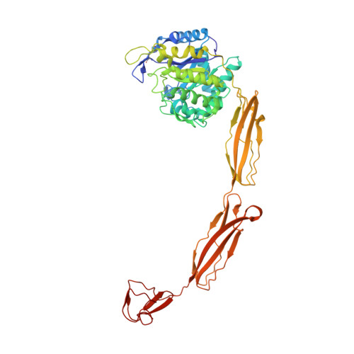
Crystal structures of substrate-bound chitinase from the psychrophilic bacterium Moritella marina and its structure in solution

Malecki, P.H.Vorgias, C.E.Petoukhov, M.V.Svergun, D.I.Rypniewski, W.

(2014) Acta Crystallogr D Biol Crystallogr 70: 676-684

  • DOI: https://doi.org/10.1107/S1399004713032264
  • Primary Citation of Related Structures:  
    4MB3, 4MB4, 4MB5

  • PubMed Abstract: 

    The four-domain structure of chitinase 60 from Moritella marina (MmChi60) is outstanding in its complexity. Many glycoside hydrolases, such as chitinases and cellulases, have multi-domain structures, but only a few have been solved. The flexibility of the hinge regions between the domains apparently makes these proteins difficult to crystallize. The analysis of an active-site mutant of MmChi60 in an unliganded form and in complex with the substrates NAG4 and NAG5 revealed significant differences in the substrate-binding site compared with the previously determined complexes of most studied chitinases. A SAXS experiment demonstrated that in addition to the elongated state found in the crystal, the protein can adapt other conformations in solution ranging from fully extended to compact.


  • Organizational Affiliation

    Institute of Bioorganic Chemistry, Polish Academy of Sciences, Noskowskiego 12/14, 61-704 Poznan, Poland.


Macromolecules
Find similar proteins by:  (by identity cutoff)  |  3D Structure
Entity ID: 1
MoleculeChains Sequence LengthOrganismDetailsImage
Chitinase 60528Moritella marinaMutation(s): 1 
Gene Names: chi60
EC: 3.2.1.14
UniProt
Find proteins for B1VBB0 (Moritella marina)
Explore B1VBB0 
Go to UniProtKB:  B1VBB0
Entity Groups  
Sequence Clusters30% Identity50% Identity70% Identity90% Identity95% Identity100% Identity
UniProt GroupB1VBB0
Sequence Annotations
Expand
  • Reference Sequence
Small Molecules
Ligands 5 Unique
IDChains Name / Formula / InChI Key2D Diagram3D Interactions
MPD
Query on MPD

Download Ideal Coordinates CCD File 
G [auth A](4S)-2-METHYL-2,4-PENTANEDIOL
C6 H14 O2
SVTBMSDMJJWYQN-YFKPBYRVSA-N
GLY
Query on GLY

Download Ideal Coordinates CCD File 
F [auth A]GLYCINE
C2 H5 N O2
DHMQDGOQFOQNFH-UHFFFAOYSA-N
IMD
Query on IMD

Download Ideal Coordinates CCD File 
H [auth A]IMIDAZOLE
C3 H5 N2
RAXXELZNTBOGNW-UHFFFAOYSA-O
CL
Query on CL

Download Ideal Coordinates CCD File 
I [auth A]CHLORIDE ION
Cl
VEXZGXHMUGYJMC-UHFFFAOYSA-M
NA
Query on NA

Download Ideal Coordinates CCD File 
B [auth A],
C [auth A],
D [auth A],
E [auth A]
SODIUM ION
Na
FKNQFGJONOIPTF-UHFFFAOYSA-N
Experimental Data & Validation

Experimental Data

  • Method: X-RAY DIFFRACTION
  • Resolution: 1.55 Å
  • R-Value Free: 0.183 
  • R-Value Work: 0.141 
  • R-Value Observed: 0.143 
  • Space Group: P 31 1 2
Unit Cell:
Length ( Å )Angle ( ˚ )
a = 67.657α = 90
b = 67.657β = 90
c = 255.643γ = 120
Software Package:
Software NamePurpose
XSCALEdata scaling
PHASERphasing
PHENIXrefinement
PDB_EXTRACTdata extraction
MAR345dtbdata collection
XDSdata reduction
XDSdata scaling

Structure Validation

View Full Validation Report


Entry History 

Deposition Data

Revision History  (Full details and data files)

  • Version 1.0: 2014-03-19
    Type: Initial release
  • Version 1.1: 2023-11-08
    Changes: Data collection, Database references, Derived calculations, Refinement description