4K5R

The 2.0 angstrom crystal structure of MTMOIV, a baeyer-villiger monooxygenase from the mithramycin biosynthetic pathway in streptomyces argillaceus.


Experimental Data Snapshot

  • Method: X-RAY DIFFRACTION
  • Resolution: 2.00 Å
  • R-Value Free: 0.249 
  • R-Value Work: 0.205 
  • R-Value Observed: 0.207 

wwPDB Validation   3D Report Full Report


Ligand Structure Quality Assessment 


This is version 1.3 of the entry. See complete history


Literature

Molecular Insight into Substrate Recognition and Catalysis of Baeyer-Villiger Monooxygenase MtmOIV, the Key Frame-Modifying Enzyme in the Biosynthesis of Anticancer Agent Mithramycin.

Bosserman, M.A.Downey, T.Noinaj, N.Buchanan, S.K.Rohr, J.

(2013) ACS Chem Biol 8: 2466-2477

  • DOI: https://doi.org/10.1021/cb400399b
  • Primary Citation of Related Structures:  
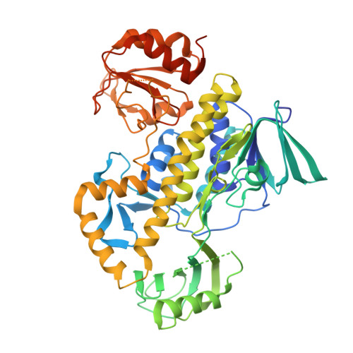
    4K5R, 4K5S

  • PubMed Abstract: 

    Baeyer-Villiger monooxygenases (BVMOs) have been shown to play key roles for the biosynthesis of important natural products. MtmOIV, a homodimeric FAD- and NADPH-dependent BVMO, catalyzes the key frame-modifying steps of the mithramycin biosynthetic pathway, including an oxidative C-C bond cleavage, by converting its natural substrate premithramycin B into mithramycin DK, the immediate precursor of mithramycin. The drastically improved protein structure of MtmOIV along with the high-resolution structure of MtmOIV in complex with its natural substrate premithramycin B are reported here, revealing previously undetected key residues that are important for substrate recognition and catalysis. Kinetic analyses of selected mutants allowed us to probe the substrate binding pocket of MtmOIV and also to discover the putative NADPH binding site. This is the first substrate-bound structure of MtmOIV providing new insights into substrate recognition and catalysis, which paves the way for the future design of a tailored enzyme for the chemo-enzymatic preparation of novel mithramycin analogues.


  • Organizational Affiliation

    Department of Pharmaceutical Sciences, College of Pharmacy, University of Kentucky, 789 South Limestone Street, Lexington, KY 40536-0596, USA.


Macromolecules
Find similar proteins by:  (by identity cutoff)  |  3D Structure
Entity ID: 1
MoleculeChains Sequence LengthOrganismDetailsImage
Oxygenase
A, B
536Streptomyces argillaceusMutation(s): 0 
Gene Names: mtmOIV
UniProt
Find proteins for Q194P4 (Streptomyces argillaceus)
Explore Q194P4 
Go to UniProtKB:  Q194P4
Entity Groups  
Sequence Clusters30% Identity50% Identity70% Identity90% Identity95% Identity100% Identity
UniProt GroupQ194P4
Sequence Annotations
Expand
  • Reference Sequence
Small Molecules
Ligands 1 Unique
IDChains Name / Formula / InChI Key2D Diagram3D Interactions
FAD
Query on FAD

Download Ideal Coordinates CCD File 
C [auth A],
D [auth B]
FLAVIN-ADENINE DINUCLEOTIDE
C27 H33 N9 O15 P2
VWWQXMAJTJZDQX-UYBVJOGSSA-N
Experimental Data & Validation

Experimental Data

  • Method: X-RAY DIFFRACTION
  • Resolution: 2.00 Å
  • R-Value Free: 0.249 
  • R-Value Work: 0.205 
  • R-Value Observed: 0.207 
  • Space Group: P 1 2 1
Unit Cell:
Length ( Å )Angle ( ˚ )
a = 83.295α = 90
b = 56.05β = 95
c = 124.903γ = 90
Software Package:
Software NamePurpose
SERGUIdata collection
PHASERphasing
PHENIXrefinement
HKL-2000data reduction
HKL-2000data scaling

Structure Validation

View Full Validation Report


Ligand Structure Quality Assessment 


Entry History 

Deposition Data

Revision History  (Full details and data files)

  • Version 1.0: 2013-10-09
    Type: Initial release
  • Version 1.1: 2013-11-27
    Changes: Database references
  • Version 1.2: 2014-11-12
    Changes: Structure summary
  • Version 1.3: 2024-02-28
    Changes: Data collection, Database references, Derived calculations