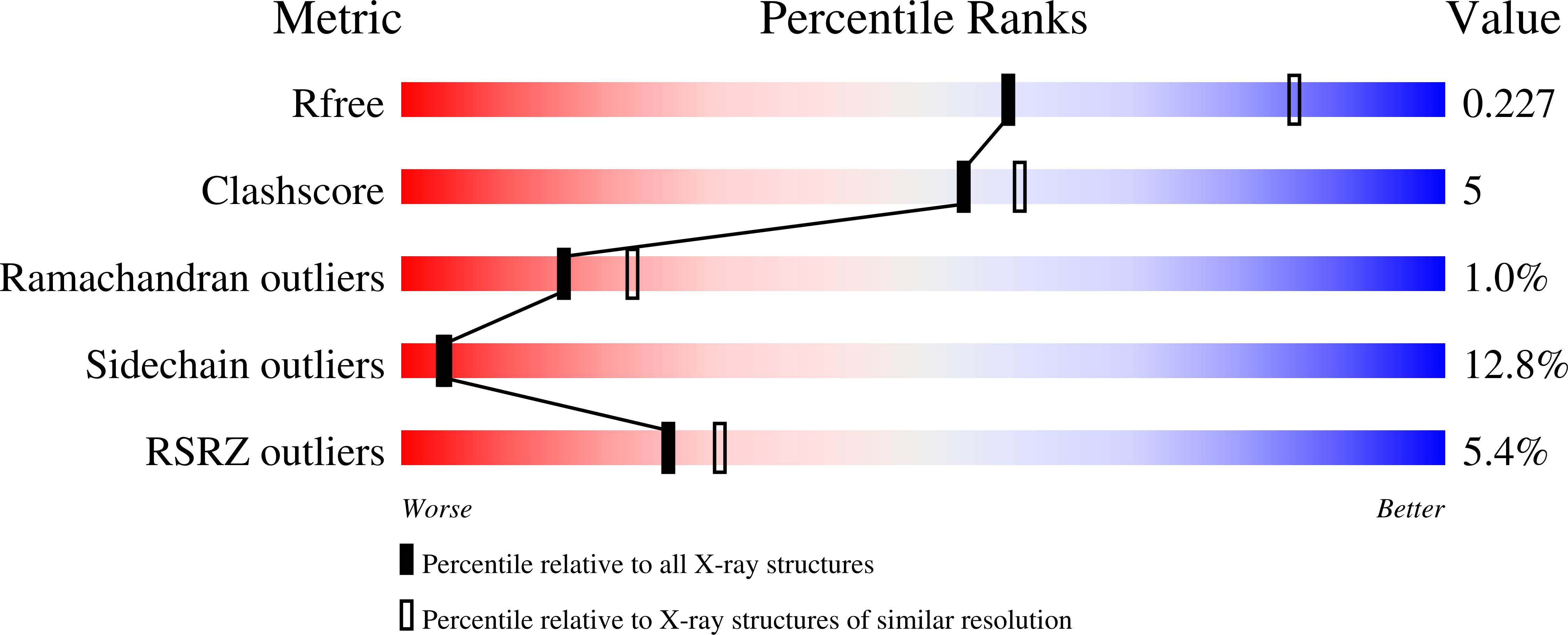
4K2X

OxyS anhydrotetracycline hydroxylase from Streptomyces rimosus


Experimental Data Snapshot

  • Method: X-RAY DIFFRACTION
  • Resolution: 2.55 Å
  • R-Value Free: 0.229 
  • R-Value Work: 0.187 
  • R-Value Observed: 0.189 

wwPDB Validation   3D Report Full Report


Ligand Structure Quality Assessment 


This is version 1.2 of the entry. See complete history


Literature

Uncovering the Enzymes that Catalyze the Final Steps in Oxytetracycline Biosynthesis.

Wang, P.Bashiri, G.Gao, X.Sawaya, M.R.Tang, Y.

(2013) J Am Chem Soc 135: 7138-7141

  • DOI: https://doi.org/10.1021/ja403516u
  • Primary Citation of Related Structures:  
    4K2X

  • PubMed Abstract: 

    Tetracyclines are a group of natural products sharing a linearly fused four-ring scaffold, which is essential for their broad-spectrum antibiotic activities. Formation of the key precursor anhydrotetracycline 3 during oxytetracycline 1 biosynthesis has been previously characterized. However, the enzymatic steps that transform 3 into 1, including the additional hydroxylation at C5 and the final C5a-C11a reduction, have remained elusive. Here we report two redox enzymes, OxyS and OxyR, are sufficient to convert 3 to 1. OxyS catalyzes two sequential hydroxylations at C6 and C5 positions of 3 with opposite stereochemistry, while OxyR catalyzes the C5a-C11a reduction using F420 as a cofactor to produce 1. The crystal structure of OxyS was obtained to provide insights into the tandem C6- and C5-hydroxylation steps. The substrate specificities of OxyS and OxyR were shown to influence the relative ratio of 1 and tetracycline 2.


  • Organizational Affiliation

    Department of Chemical and Biomolecular Engineering, University of California Los Angeles, Los Angeles, California 90095, USA.


Macromolecules
Find similar proteins by:  (by identity cutoff)  |  3D Structure
Entity ID: 1
MoleculeChains Sequence LengthOrganismDetailsImage
Polyketide oxygenase/hydroxylase
A, B
523Streptomyces rimosus subsp. rimosus ATCC 10970Mutation(s): 0 
Gene Names: oxySSRIM_10936
UniProt
Find proteins for L8EUQ6 (Streptomyces rimosus subsp. rimosus (strain ATCC 10970 / DSM 40260 / JCM 4667 / NRRL 2234))
Explore L8EUQ6 
Go to UniProtKB:  L8EUQ6
Entity Groups  
Sequence Clusters30% Identity50% Identity70% Identity90% Identity95% Identity100% Identity
UniProt GroupL8EUQ6
Sequence Annotations
Expand
  • Reference Sequence
Small Molecules
Ligands 1 Unique
IDChains Name / Formula / InChI Key2D Diagram3D Interactions
FAD
Query on FAD

Download Ideal Coordinates CCD File 
C [auth A],
D [auth B]
FLAVIN-ADENINE DINUCLEOTIDE
C27 H33 N9 O15 P2
VWWQXMAJTJZDQX-UYBVJOGSSA-N
Experimental Data & Validation

Experimental Data

  • Method: X-RAY DIFFRACTION
  • Resolution: 2.55 Å
  • R-Value Free: 0.229 
  • R-Value Work: 0.187 
  • R-Value Observed: 0.189 
  • Space Group: P 21 21 21
Unit Cell:
Length ( Å )Angle ( ˚ )
a = 76.06α = 90
b = 115.13β = 90
c = 121.03γ = 90
Software Package:
Software NamePurpose
XSCALEdata scaling
PHASERphasing
BUSTER-TNTrefinement
PDB_EXTRACTdata extraction
ADSCdata collection
BUSTERrefinement

Structure Validation

View Full Validation Report


Ligand Structure Quality Assessment 


Entry History 

Deposition Data

Revision History  (Full details and data files)

  • Version 1.0: 2013-05-15
    Type: Initial release
  • Version 1.1: 2013-06-19
    Changes: Database references
  • Version 1.2: 2023-09-20
    Changes: Data collection, Database references, Derived calculations, Refinement description