4JD0

Structure of the inositol-1-phosphate CTP transferase from T. maritima.


Experimental Data Snapshot

  • Method: X-RAY DIFFRACTION
  • Resolution: 1.80 Å
  • R-Value Free: 0.166 
  • R-Value Work: 0.111 
  • R-Value Observed: 0.114 

wwPDB Validation   3D Report Full Report


Ligand Structure Quality Assessment 


This is version 1.1 of the entry. See complete history


Literature

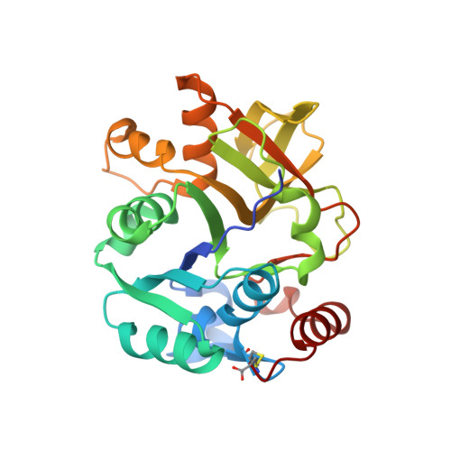
Structure of the inositol-1-phosphate cytidylyltransferase from Thermotoga maritima.

Kurnasov, O.V.Luk, H.J.Roberts, M.F.Stec, B.

(2013) Acta Crystallogr D Biol Crystallogr 69: 1808-1817

  • DOI: https://doi.org/10.1107/S0907444913015278
  • Primary Citation of Related Structures:  
    4JD0

  • PubMed Abstract: 

    The unique steps in the synthesis of an unusual osmolyte in hyperthermophiles, di-myo-inositol-1,1'-phosphate (DIP), involve the production of CDP-inositol and its condensation with an inositol-1-phosphate molecule to form phosphorylated DIP. While many organisms fuse both activities into a single enzyme, the two are separate in Thermotoga maritima. The crystal structure of the T. maritima inositol-1-phosphate cytidylyltransferase, which as a soluble protein may transiently associate with its membrane-embedded partner phospho-DIP synthase (P-DIPS), has now been obtained. The structure shows a conserved motif of sugar nucleotide transferases (COG1213) with a structurally reinforced C-terminal Cys bonded to the core of the protein. A bound arsenosugar identifies the location of the active site for inositol 1-phosphate. Based on homologous structures from several species and the identification of the crucial conserved aspartate residue, a catalytic mechanism for this enzyme is proposed as well as a mode for its association with P-DIPS. This structure imposes constraints on the mode of association, communication and temperature activation of two separate enzymes in T. maritima. For the first time, a working model for the membrane-bound P-DIPS unit has been constructed. This sheds light on the functioning of the phosphatidylserine and phosphatidylinositol synthases involved in many physiological processes that are homologous to P-DIPS. This work provides fresh insights into the synthesis of the unusual thermoprotective compound DIP in hyperthermophiles.


  • Organizational Affiliation

    Sanford-Burnham Institute for Medical Research, La Jolla, CA 92037, USA.


Macromolecules
Find similar proteins by:  (by identity cutoff)  |  3D Structure
Entity ID: 1
MoleculeChains Sequence LengthOrganismDetailsImage
Nucleotidyl transferase251Thermotoga maritima MSB8Mutation(s): 0 
Gene Names: ThemaDRAFT_0946TM1418A
Entity Groups  
Sequence Clusters30% Identity50% Identity70% Identity90% Identity95% Identity100% Identity
Sequence Annotations
Expand
  • Reference Sequence
Small Molecules
Ligands 5 Unique
IDChains Name / Formula / InChI Key2D Diagram3D Interactions
1KH
Query on 1KH

Download Ideal Coordinates CCD File 
B [auth A]arsenoribose
C5 H13 As O8
MSQKZAOQSOMILW-FLRLBIABSA-N
PO4
Query on PO4

Download Ideal Coordinates CCD File 
E [auth A],
F [auth A],
G [auth A]
PHOSPHATE ION
O4 P
NBIIXXVUZAFLBC-UHFFFAOYSA-K
GOL
Query on GOL

Download Ideal Coordinates CCD File 
N [auth A],
O [auth A]
GLYCEROL
C3 H8 O3
PEDCQBHIVMGVHV-UHFFFAOYSA-N
CL
Query on CL

Download Ideal Coordinates CCD File 
C [auth A]CHLORIDE ION
Cl
VEXZGXHMUGYJMC-UHFFFAOYSA-M
NA
Query on NA

Download Ideal Coordinates CCD File 
D [auth A]
H [auth A]
I [auth A]
J [auth A]
K [auth A]
D [auth A],
H [auth A],
I [auth A],
J [auth A],
K [auth A],
L [auth A],
M [auth A],
P [auth A]
SODIUM ION
Na
FKNQFGJONOIPTF-UHFFFAOYSA-N
Experimental Data & Validation

Experimental Data

  • Method: X-RAY DIFFRACTION
  • Resolution: 1.80 Å
  • R-Value Free: 0.166 
  • R-Value Work: 0.111 
  • R-Value Observed: 0.114 
  • Space Group: P 32 2 1
Unit Cell:
Length ( Å )Angle ( ˚ )
a = 133.09α = 90
b = 133.09β = 90
c = 43.365γ = 120
Software Package:
Software NamePurpose
CrystalCleardata collection
PHASESphasing
REFMACrefinement
HKL-2000data reduction
SCALEPACKdata scaling

Structure Validation

View Full Validation Report


Ligand Structure Quality Assessment 


Entry History 

Deposition Data

  • Released Date: 2013-09-18 
  • Deposition Author(s): Stec, B.

Revision History  (Full details and data files)

  • Version 1.0: 2013-09-18
    Type: Initial release
  • Version 1.1: 2023-09-20
    Changes: Data collection, Database references, Derived calculations, Refinement description