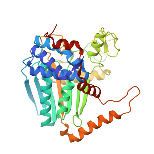
Structural and functional insights into (s)-ureidoglycolate dehydrogenase, a metabolic branch point enzyme in nitrogen utilization.
Kim, M.I., Shin, I., Cho, S., Lee, J., Rhee, S.(2012) PLoS One 7: e52066-e52066
- PubMed: 23284870
- DOI: https://doi.org/10.1371/journal.pone.0052066
- Primary Citation of Related Structures:
4FJS, 4FJU, 4H8A - PubMed Abstract:
Nitrogen metabolism is one of essential processes in living organisms. The catabolic pathways of nitrogenous compounds play a pivotal role in the storage and recovery of nitrogen. In Escherichia coli, two different, interconnecting metabolic routes drive nitrogen utilization through purine degradation metabolites. The enzyme (S)-ureidoglycolate dehydrogenase (AllD), which is a member of l-sulfolactate dehydrogenase-like family, converts (S)-ureidoglycolate, a key intermediate in the purine degradation pathway, to oxalurate in an NAD(P)-dependent manner. Therefore, AllD is a metabolic branch-point enzyme for nitrogen metabolism in E. coli. Here, we report crystal structures of AllD in its apo form, in a binary complex with NADH cofactor, and in a ternary complex with NADH and glyoxylate, a possible spontaneous degradation product of oxalurate. Structural analyses revealed that NADH in an extended conformation is bound to an NADH-binding fold with three distinct domains that differ from those of the canonical NADH-binding fold. We also characterized ligand-induced structural changes, as well as the binding mode of glyoxylate, in the active site near the NADH nicotinamide ring. Based on structural and kinetic analyses, we concluded that AllD selectively utilizes NAD(+) as a cofactor, and further propose that His116 acts as a general catalytic base and that a hydride transfer is possible on the B-face of the nicotinamide ring of the cofactor. Other residues conserved in the active sites of this novel l-sulfolactate dehydrogenase-like family also play essential roles in catalysis.
Organizational Affiliation:
Department of Agricultural Biotechnology, Seoul National University, Seoul, Korea.