4DXT

Human SUN2 (AA 522-717)


Experimental Data Snapshot

  • Method: X-RAY DIFFRACTION
  • Resolution: 2.22 Å
  • R-Value Free: 0.226 
  • R-Value Work: 0.169 
  • R-Value Observed: 0.175 

wwPDB Validation   3D Report Full Report


This is version 1.0 of the entry. See complete history


Literature

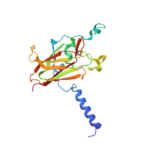
LINC Complexes Form by Binding of Three KASH Peptides to Domain Interfaces of Trimeric SUN Proteins.

Sosa, B.A.Rothballer, A.Kutay, U.Schwartz, T.U.

(2012) Cell 149: 1035-1047

  • DOI: https://doi.org/10.1016/j.cell.2012.03.046
  • Primary Citation of Related Structures:  
    4DXR, 4DXS, 4DXT

  • PubMed Abstract: 

    Linker of nucleoskeleton and cytoskeleton (LINC) complexes span the nuclear envelope and are composed of KASH and SUN proteins residing in the outer and inner nuclear membrane, respectively. LINC formation relies on direct binding of KASH and SUN in the perinuclear space. Thereby, molecular tethers are formed that can transmit forces for chromosome movements, nuclear migration, and anchorage. We present crystal structures of the human SUN2-KASH1/2 complex, the core of the LINC complex. The SUN2 domain is rigidly attached to a trimeric coiled coil that prepositions it to bind three KASH peptides. The peptides bind in three deep and expansive grooves formed between adjacent SUN domains, effectively acting as molecular glue. In addition, a disulfide between conserved cysteines on SUN and KASH covalently links both proteins. The structure provides the basis of LINC complex formation and suggests a model for how LINC complexes might arrange into higher-order clusters to enhance force-coupling.


  • Organizational Affiliation

    Department of Biology, Massachusetts Institute of Technology, 77 Massachusetts Avenue, Cambridge, MA 02139, USA.


Macromolecules
Find similar proteins by:  (by identity cutoff)  |  3D Structure
Entity ID: 1
MoleculeChains Sequence LengthOrganismDetailsImage
SUN domain-containing protein 2198Homo sapiensMutation(s): 0 
Gene Names: SUN2FRIGGKIAA0668RAB5IPUNC84B
UniProt & NIH Common Fund Data Resources
Find proteins for Q9UH99 (Homo sapiens)
Explore Q9UH99 
Go to UniProtKB:  Q9UH99
PHAROS:  Q9UH99
GTEx:  ENSG00000100242 
Entity Groups  
Sequence Clusters30% Identity50% Identity70% Identity90% Identity95% Identity100% Identity
UniProt GroupQ9UH99
Sequence Annotations
Expand
  • Reference Sequence
Small Molecules
Ligands 1 Unique
IDChains Name / Formula / InChI Key2D Diagram3D Interactions
K
Query on K

Download Ideal Coordinates CCD File 
B [auth A]POTASSIUM ION
K
NPYPAHLBTDXSSS-UHFFFAOYSA-N
Experimental Data & Validation

Experimental Data

  • Method: X-RAY DIFFRACTION
  • Resolution: 2.22 Å
  • R-Value Free: 0.226 
  • R-Value Work: 0.169 
  • R-Value Observed: 0.175 
  • Space Group: H 3 2
Unit Cell:
Length ( Å )Angle ( ˚ )
a = 79.58α = 90
b = 79.58β = 90
c = 199.05γ = 120
Software Package:
Software NamePurpose
ADSCdata collection
PHENIXrefinement
HKL-2000data scaling

Structure Validation

View Full Validation Report


Entry History 

Deposition Data

Revision History  (Full details and data files)

  • Version 1.0: 2012-06-06
    Type: Initial release