4DDJ

Crystal structure of saposin A in complex with lauryldimethylamine-N-oxide (LDAO)


Experimental Data Snapshot

  • Method: X-RAY DIFFRACTION
  • Resolution: 1.90 Å
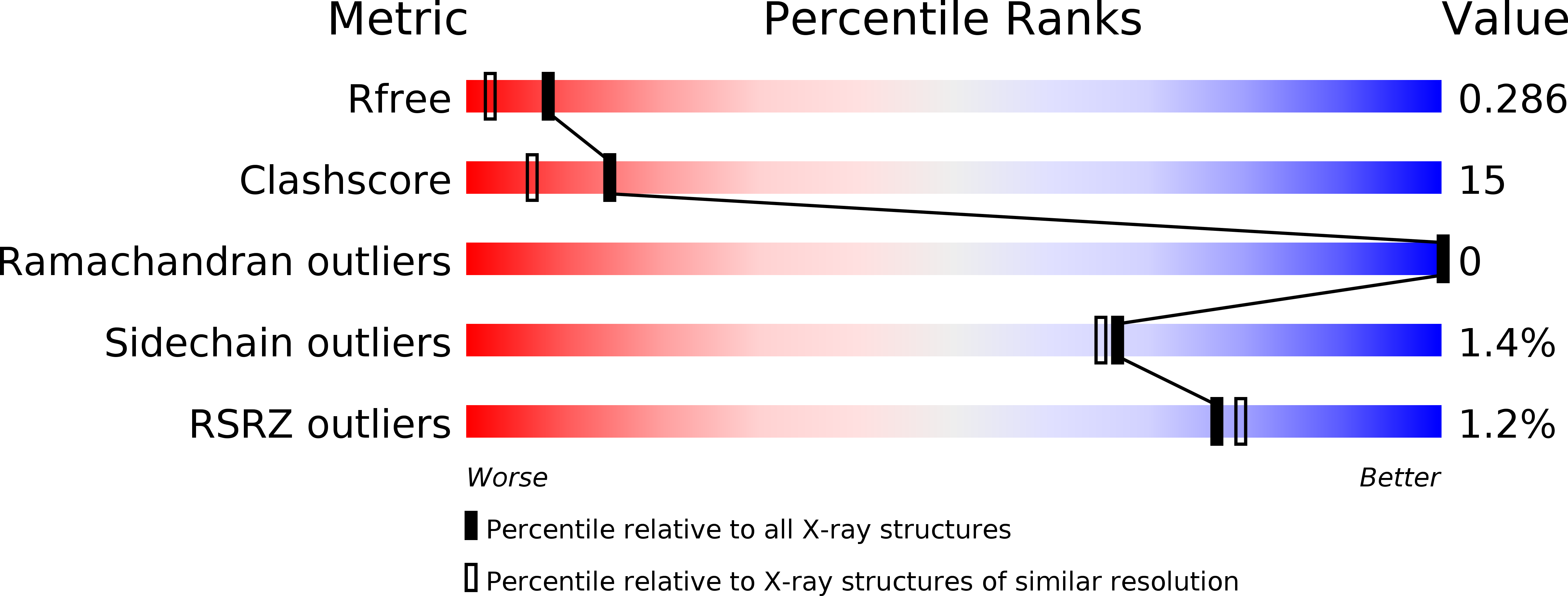
  • R-Value Free: 0.280 
  • R-Value Work: 0.230 
  • R-Value Observed: 0.232 

wwPDB Validation   3D Report Full Report


Ligand Structure Quality Assessment 


This is version 1.3 of the entry. See complete history


Literature

Structure of saposin A lipoprotein discs.

Popovic, K.Holyoake, J.Pomes, R.Prive, G.G.

(2012) Proc Natl Acad Sci U S A 109: 2908-2912

  • DOI: https://doi.org/10.1073/pnas.1115743109
  • Primary Citation of Related Structures:  
    4DDJ

  • PubMed Abstract: 

    The saposins are small, membrane-active proteins that exist in both soluble and lipid-bound states. Saposin A has roles in sphingolipid catabolism and transport and is required for the breakdown of galactosylceramide by β-galactosylceramidase. In the absence of lipid, saposin A adopts a closed monomeric apo conformation typical of this family. To study a lipid-bound state of this protein, we determined the crystal structure of saposin A in the presence of detergent to 1.9 Å resolution. The structure reveals two chains of saposin A in an open conformation encapsulating 40 internally bound detergent molecules organized in a highly ordered bilayer-like hydrophobic core. The complex provides a high-resolution view of a discoidal lipoprotein particle in which all of the internalized acyl chains are resolved. Saposin A lipoprotein discs exhibit limited selectivity with respect to the incorporated lipid, and can solubilize phospholipids, sphingolipids, and cholesterol into discrete, monodisperse particles with mass of approximately 27 kDa. These discs may be the smallest possible lipoprotein structures that are stabilized by lipid self-assembly.


  • Organizational Affiliation

    Department of Medical Biophysics, University of Toronto, Toronto, ON, Canada M5G 2M9.


Macromolecules
Find similar proteins by:  (by identity cutoff)  |  3D Structure
Entity ID: 1
MoleculeChains Sequence LengthOrganismDetailsImage
Saposin-A83Homo sapiensMutation(s): 0 
Gene Names: PSAPGLBASAP1
Membrane Entity: Yes 
UniProt & NIH Common Fund Data Resources
Find proteins for P07602 (Homo sapiens)
Explore P07602 
Go to UniProtKB:  P07602
PHAROS:  P07602
GTEx:  ENSG00000197746 
Entity Groups  
Sequence Clusters30% Identity50% Identity70% Identity90% Identity95% Identity100% Identity
UniProt GroupP07602
Sequence Annotations
Expand
  • Reference Sequence
Small Molecules
Ligands 1 Unique
IDChains Name / Formula / InChI Key2D Diagram3D Interactions
LDA
Query on LDA

Download Ideal Coordinates CCD File 
B [auth A]
C [auth A]
D [auth A]
E [auth A]
F [auth A]
B [auth A],
C [auth A],
D [auth A],
E [auth A],
F [auth A],
G [auth A],
H [auth A],
I [auth A],
J [auth A],
K [auth A],
L [auth A],
M [auth A],
N [auth A],
O [auth A],
P [auth A],
Q [auth A],
R [auth A],
S [auth A],
T [auth A],
U [auth A]
LAURYL DIMETHYLAMINE-N-OXIDE
C14 H31 N O
SYELZBGXAIXKHU-UHFFFAOYSA-N
Experimental Data & Validation

Experimental Data

  • Method: X-RAY DIFFRACTION
  • Resolution: 1.90 Å
  • R-Value Free: 0.280 
  • R-Value Work: 0.230 
  • R-Value Observed: 0.232 
  • Space Group: P 61 2 2
Unit Cell:
Length ( Å )Angle ( ˚ )
a = 39.794α = 90
b = 39.794β = 90
c = 247.284γ = 120
Software Package:
Software NamePurpose
REFMACrefinement
PDB_EXTRACTdata extraction
HKL-3000data collection
HKL-3000data reduction
HKL-3000data scaling
SHELXSphasing

Structure Validation

View Full Validation Report


Ligand Structure Quality Assessment 


Entry History 

Deposition Data

Revision History  (Full details and data files)

  • Version 1.0: 2012-02-15
    Type: Initial release
  • Version 1.1: 2012-02-22
    Changes: Database references
  • Version 1.2: 2012-03-14
    Changes: Database references
  • Version 1.3: 2020-01-29
    Changes: Advisory, Database references, Derived calculations