4BWF

Pex4p-Pex22p disulphide bond mutant


Experimental Data Snapshot

  • Method: X-RAY DIFFRACTION
  • Resolution: 3.23 Å
  • R-Value Free: 0.264 
  • R-Value Work: 0.203 
  • R-Value Observed: 0.205 

wwPDB Validation   3D Report Full Report


This is version 1.4 of the entry. See complete history


Literature

A Disulphide Bond in the E2 Enzyme Pex4P Modulates Ubiquitin-Conjugating Activity

Williams, C.van den Berg, M.Stanley, W.A.Wilmanns, M.Distel, B.

(2013) Sci Rep 3: 2212

  • DOI: https://doi.org/10.1038/srep02212
  • Primary Citation of Related Structures:  
    4BWF

  • PubMed Abstract: 

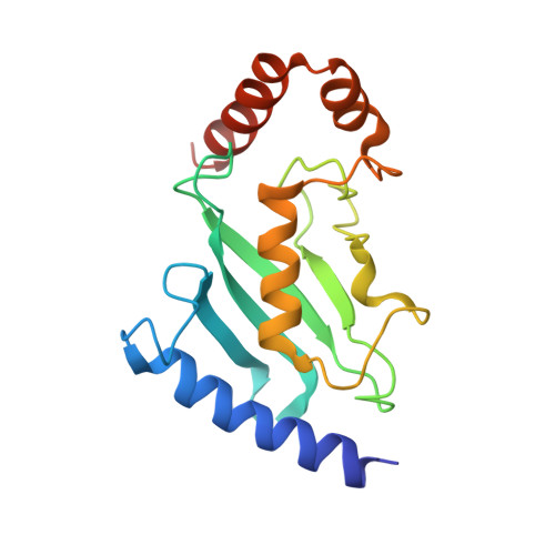
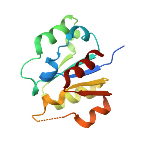
    The ubiquitin-conjugating enzyme Pex4p together with its binding partner, the peroxisomal membrane protein Pex22p, co-ordinates cysteine-dependent ubiquitination of the cycling receptor protein Pex5p. Unusually for an ubiquitin-conjugating enzyme, Saccharomyces cerevisiae Pex4p can form a disulphide bond between the cysteine residues at positions 105 and 146. We found that mutating the disulphide forming cysteine residues in Pex4p to serines does not disturb the secondary structure of the protein but does reduce the in vitro activity of Pex4p. From the crystal structure of Pex4p C105S, C146S in complex with the soluble domain of Pex22p, we observe a narrowing of the active site cleft, caused by loss of the disulphide bond. This modification of the active site microenvironment is likely to restrict access of ubiquitin to the active site cysteine, modulating Pex4p activity. Finally, based on sequence and structural alignments, we have identified other ubiquitin-conjugating enzymes that may contain disulphide bonds.


  • Organizational Affiliation

    European Molecular Biology Laboratory, Structural Biology Unit, Notkestrasse 85, 22603, Hamburg, Germany.


Macromolecules
Find similar proteins by:  (by identity cutoff)  |  3D Structure
Entity ID: 1
MoleculeChains Sequence LengthOrganismDetailsImage
UBIQUITIN-CONJUGATING ENZYME E2-21 KDA172Saccharomyces cerevisiaeMutation(s): 3 
EC: 6.3.2.19
UniProt
Find proteins for P29340 (Saccharomyces cerevisiae (strain ATCC 204508 / S288c))
Explore P29340 
Go to UniProtKB:  P29340
Entity Groups  
Sequence Clusters30% Identity50% Identity70% Identity90% Identity95% Identity100% Identity
UniProt GroupP29340
Sequence Annotations
Expand
  • Reference Sequence
Find similar proteins by:  (by identity cutoff)  |  3D Structure
Entity ID: 2
MoleculeChains Sequence LengthOrganismDetailsImage
PEROXISOME ASSEMBLY PROTEIN 22130Saccharomyces cerevisiaeMutation(s): 0 
UniProt
Find proteins for P39718 (Saccharomyces cerevisiae (strain ATCC 204508 / S288c))
Explore P39718 
Go to UniProtKB:  P39718
Entity Groups  
Sequence Clusters30% Identity50% Identity70% Identity90% Identity95% Identity100% Identity
UniProt GroupP39718
Sequence Annotations
Expand
  • Reference Sequence
Experimental Data & Validation

Experimental Data

  • Method: X-RAY DIFFRACTION
  • Resolution: 3.23 Å
  • R-Value Free: 0.264 
  • R-Value Work: 0.203 
  • R-Value Observed: 0.205 
  • Space Group: C 1 2 1
Unit Cell:
Length ( Å )Angle ( ˚ )
a = 138.858α = 90
b = 42.802β = 100.14
c = 60.54γ = 90
Software Package:
Software NamePurpose
REFMACrefinement
XDSdata reduction
SCALAdata scaling
MOLREPphasing

Structure Validation

View Full Validation Report


Entry History 

Deposition Data

Revision History  (Full details and data files)

  • Version 1.0: 2013-07-17
    Type: Initial release
  • Version 1.1: 2013-08-07
    Changes: Database references
  • Version 1.2: 2017-12-13
    Changes: Database references
  • Version 1.3: 2019-07-17
    Changes: Data collection
  • Version 1.4: 2023-12-20
    Changes: Data collection, Database references, Derived calculations, Other, Refinement description