4B9Z

Crystal Structure of Agd31B, alpha-transglucosylase, complexed with Acarbose


Experimental Data Snapshot

  • Method: X-RAY DIFFRACTION
  • Resolution: 2.00 Å
  • R-Value Free: 0.187 
  • R-Value Work: 0.161 
  • R-Value Observed: 0.162 

wwPDB Validation   3D Report Full Report


This is version 2.1 of the entry. See complete history


Literature

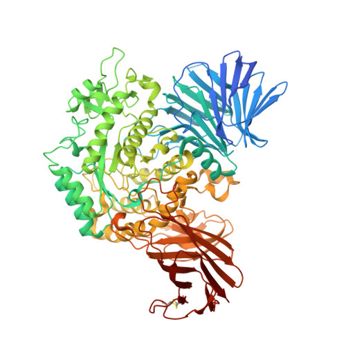
Structural Enzymology of Cellvibrio Japonicus Agd31B Reveals Alpha-Transglucosylase Activity in Glycoside Hydrolase Family 31

Larsbrink, J.Izumi, A.Hemsworth, G.R.Davies, G.J.Brumer, H.

(2012) J Biol Chem 287: 43288

  • DOI: https://doi.org/10.1074/jbc.M112.416511
  • Primary Citation of Related Structures:  
    4B9Y, 4B9Z, 4BA0

  • PubMed Abstract: 

    The metabolism of the storage polysaccharides glycogen and starch is of vital importance to organisms from all domains of life. In bacteria, utilization of these α-glucans requires the concerted action of a variety of enzymes, including glycoside hydrolases, glycoside phosphorylases, and transglycosylases. In particular, transglycosylases from glycoside hydrolase family 13 (GH13) and GH77 play well established roles in α-glucan side chain (de)branching, regulation of oligo- and polysaccharide chain length, and formation of cyclic dextrans. Here, we present the biochemical and tertiary structural characterization of a new type of bacterial 1,4-α-glucan 4-α-glucosyltransferase from GH31. Distinct from 1,4-α-glucan 6-α-glucosyltransferases (EC 2.4.1.24) and 4-α-glucanotransferases (EC 2.4.1.25), this enzyme strictly transferred one glucosyl residue from α(1→4)-glucans in disproportionation reactions. Substrate hydrolysis was undetectable for a series of malto-oligosaccharides except maltose for which transglycosylation nonetheless dominated across a range of substrate concentrations. Crystallographic analysis of the enzyme in free, acarbose-complexed, and trapped 5-fluoro-β-glucosyl-enzyme intermediate forms revealed extended substrate interactions across one negative and up to three positive subsites, thus providing structural rationalization for the unique, single monosaccharide transferase activity of the enzyme.


  • Organizational Affiliation

    Division of Glycoscience, School of Biotechnology, Royal Institute of Technology, AlbaNova University Centre, 106 91 Stockholm, Sweden.


Macromolecules
Find similar proteins by:  (by identity cutoff)  |  3D Structure
Entity ID: 1
MoleculeChains Sequence LengthOrganismDetailsImage
ALPHA-GLUCOSIDASE, PUTATIVE, ADG31B817Cellvibrio japonicusMutation(s): 0 
EC: 3.2.1.20
UniProt
Find proteins for B3PEE6 (Cellvibrio japonicus (strain Ueda107))
Explore B3PEE6 
Go to UniProtKB:  B3PEE6
Entity Groups  
Sequence Clusters30% Identity50% Identity70% Identity90% Identity95% Identity100% Identity
UniProt GroupB3PEE6
Sequence Annotations
Expand
  • Reference Sequence
Oligosaccharides

Help

Entity ID: 2
MoleculeChains Length2D Diagram Glycosylation3D Interactions
4,6-dideoxy-4-{[(1S,4R,5S,6S)-4,5,6-trihydroxy-3-(hydroxymethyl)cyclohex-2-en-1-yl]amino}-alpha-D-glucopyranose-(1-4)-alpha-D-glucopyranose-(1-4)-alpha-D-glucopyranose
B
3N/AN/A
Glycosylation Resources
GlyTouCan:  G66431MI
GlyCosmos:  G66431MI
Small Molecules
Ligands 4 Unique
IDChains Name / Formula / InChI Key2D Diagram3D Interactions
PEG
Query on PEG

Download Ideal Coordinates CCD File 
I [auth A]DI(HYDROXYETHYL)ETHER
C4 H10 O3
MTHSVFCYNBDYFN-UHFFFAOYSA-N
SO4
Query on SO4

Download Ideal Coordinates CCD File 
C [auth A],
D [auth A],
E [auth A],
F [auth A],
G [auth A]
SULFATE ION
O4 S
QAOWNCQODCNURD-UHFFFAOYSA-L
OXL
Query on OXL

Download Ideal Coordinates CCD File 
H [auth A]OXALATE ION
C2 O4
MUBZPKHOEPUJKR-UHFFFAOYSA-L
EDO
Query on EDO

Download Ideal Coordinates CCD File 
J [auth A]
K [auth A]
L [auth A]
M [auth A]
N [auth A]
J [auth A],
K [auth A],
L [auth A],
M [auth A],
N [auth A],
O [auth A],
P [auth A],
Q [auth A],
R [auth A],
S [auth A],
T [auth A],
U [auth A],
V [auth A],
W [auth A],
X [auth A]
1,2-ETHANEDIOL
C2 H6 O2
LYCAIKOWRPUZTN-UHFFFAOYSA-N
Biologically Interesting Molecules (External Reference) 1 Unique
Experimental Data & Validation

Experimental Data

  • Method: X-RAY DIFFRACTION
  • Resolution: 2.00 Å
  • R-Value Free: 0.187 
  • R-Value Work: 0.161 
  • R-Value Observed: 0.162 
  • Space Group: P 6 2 2
Unit Cell:
Length ( Å )Angle ( ˚ )
a = 196.88α = 90
b = 196.88β = 90
c = 102.76γ = 120
Software Package:
Software NamePurpose
PHENIXrefinement
MOSFLMdata reduction
SCALEPACKdata scaling
MOLREPphasing

Structure Validation

View Full Validation Report


Entry History 

Deposition Data

Revision History  (Full details and data files)

  • Version 1.0: 2012-11-14
    Type: Initial release
  • Version 1.1: 2012-11-21
    Changes: Database references
  • Version 1.2: 2013-01-16
    Changes: Database references
  • Version 2.0: 2020-07-29
    Type: Remediation
    Reason: Carbohydrate remediation
    Changes: Advisory, Atomic model, Data collection, Derived calculations, Non-polymer description, Other, Structure summary
  • Version 2.1: 2023-12-20
    Changes: Advisory, Data collection, Database references, Refinement description, Structure summary