4B0F

Heptameric core complex structure of C4b-binding (C4BP) protein from human


Experimental Data Snapshot

  • Method: X-RAY DIFFRACTION
  • Resolution: 2.80 Å
  • R-Value Free: 0.248 
  • R-Value Work: 0.212 
  • R-Value Observed: 0.214 

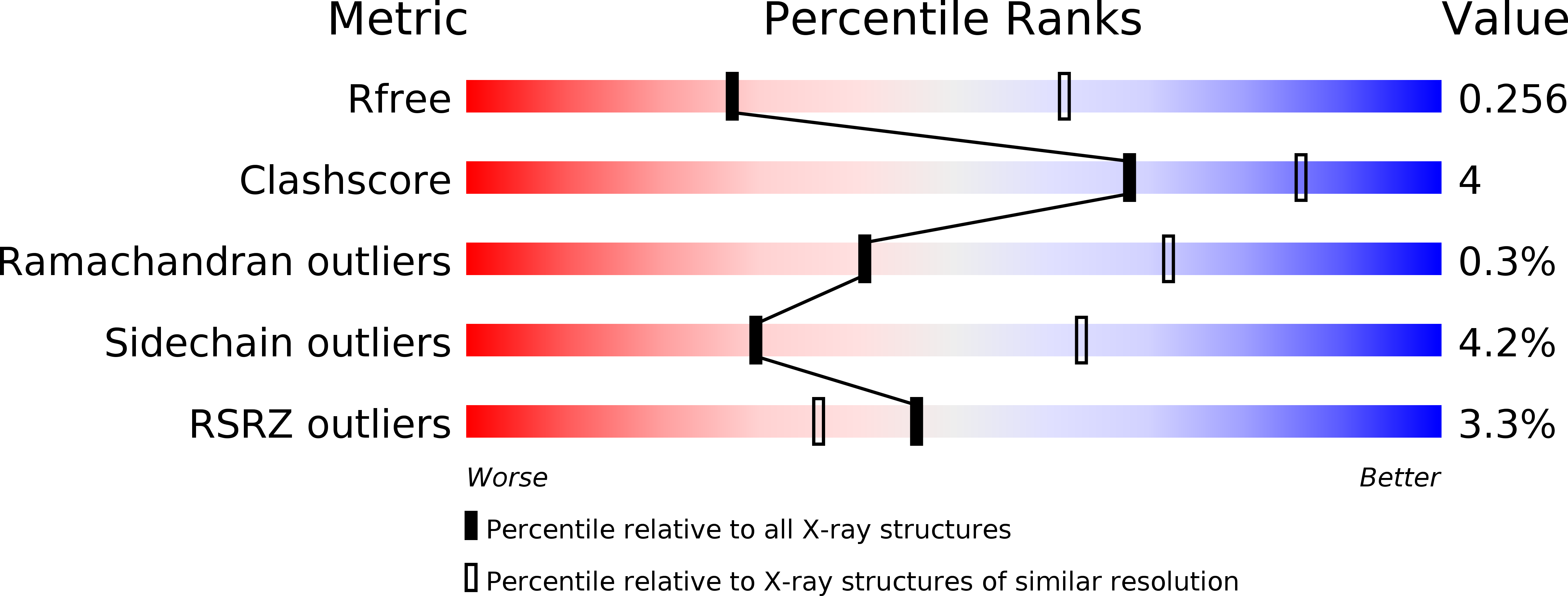
wwPDB Validation   3D Report Full Report


This is version 1.2 of the entry. See complete history


Literature

Arranged Sevenfold: Structural Insights Into the C-Terminal Oligomerization Domain of Human C4B-Binding Protein.

Hofmeyer, T.Schmelz, S.Degiacomi, M.T.Peraro, M.D.Daneschdar, M.Scrima, A.Den Heuvel, J.V.Heinz, D.W.Kolmar, H.

(2013) J Mol Biol 425: 1302

  • DOI: https://doi.org/10.1016/j.jmb.2012.12.017
  • Primary Citation of Related Structures:  
    4B0F

  • PubMed Abstract: 

    The complement system as a major part of innate immunity is the first line of defense against invading microorganisms. Orchestrated by more than 60 proteins, its major task is to discriminate between host cells and pathogens and to initiate immune response. Additional recognition of necrotic or apoptotic cells demands a fine-tune regulation of this powerful system. C4b-binding protein (C4BP) is the major inhibitor of the classical complement and lectin pathway. The crystal structure of the human C4BP oligomerization domain in its 7α isoform and molecular simulations provide first structural insights of C4BP oligomerization. The heptameric core structure is stabilized by intermolecular disulfide bonds. In addition, thermal shift assays indicate that layers of electrostatic interactions mainly contribute to the extraordinary thermodynamic stability of the complex. These findings make C4BP a promising scaffold for multivalent ligand display with applications in immunology and biological chemistry.


  • Organizational Affiliation

    Institute for Organic Chemistry and Biochemistry, Technische Universität Darmstadt, Petersenstraße 22, 64287 Darmstadt, Germany.


Macromolecules
Find similar proteins by:  (by identity cutoff)  |  3D Structure
Entity ID: 1
MoleculeChains Sequence LengthOrganismDetailsImage
C4B-BINDING PROTEIN ALPHA CHAIN
A, B, C, D, E
A, B, C, D, E, F, G
65Homo sapiensMutation(s): 0 
UniProt & NIH Common Fund Data Resources
Find proteins for P04003 (Homo sapiens)
Explore P04003 
Go to UniProtKB:  P04003
PHAROS:  P04003
GTEx:  ENSG00000123838 
Entity Groups  
Sequence Clusters30% Identity50% Identity70% Identity90% Identity95% Identity100% Identity
UniProt GroupP04003
Sequence Annotations
Expand
  • Reference Sequence
Small Molecules
Modified Residues  1 Unique
IDChains TypeFormula2D DiagramParent
MSE
Query on MSE
A, B, C, D, E
A, B, C, D, E, F, G
L-PEPTIDE LINKINGC5 H11 N O2 SeMET
Experimental Data & Validation

Experimental Data

  • Method: X-RAY DIFFRACTION
  • Resolution: 2.80 Å
  • R-Value Free: 0.248 
  • R-Value Work: 0.212 
  • R-Value Observed: 0.214 
  • Space Group: P 43 21 2
Unit Cell:
Length ( Å )Angle ( ˚ )
a = 73.85α = 90
b = 73.85β = 90
c = 204.29γ = 90
Software Package:
Software NamePurpose
PHENIXrefinement
XDSdata reduction
XSCALEdata scaling
PHENIXphasing

Structure Validation

View Full Validation Report


Entry History 

Deposition Data

Revision History  (Full details and data files)

  • Version 1.0: 2013-01-09
    Type: Initial release
  • Version 1.1: 2013-04-10
    Changes: Database references
  • Version 1.2: 2017-07-12
    Changes: Refinement description