4AIO

Crystal structure of the starch debranching enzyme barley limit dextrinase


Experimental Data Snapshot

  • Method: X-RAY DIFFRACTION
  • Resolution: 1.90 Å
  • R-Value Free: 0.225 
  • R-Value Work: 0.187 
  • R-Value Observed: 0.189 

wwPDB Validation   3D Report Full Report


This is version 1.4 of the entry. See complete history


Literature

Structure of the Starch-Debranching Enzyme Barley Limit Dextrinase Reveals Homology of the N-Terminal Domain to Cbm21.

Moeller, M.S.Abou Hachem, M.Svensson, B.Henriksen, A.

(2012) Acta Crystallogr Sect F Struct Biol Cryst Commun 68: 1008

  • DOI: https://doi.org/10.1107/S1744309112031004
  • Primary Citation of Related Structures:  
    4AIO

  • PubMed Abstract: 

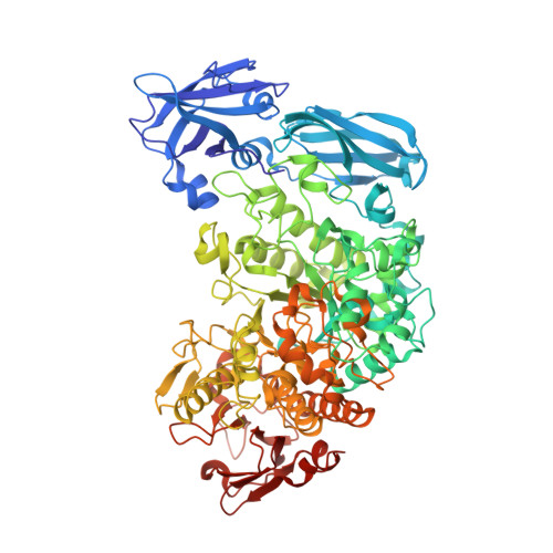
    Barley limit dextrinase (HvLD) is a debranching enzyme from glycoside hydrolase family 13 subfamily 13 (GH13_13) that hydrolyses α-1,6-glucosidic linkages in limit dextrins derived from amylopectin. The structure of HvLD was solved and refined to 1.9 Å resolution. The structure has a glycerol molecule in the active site and is virtually identical to the structures of HvLD in complex with the competitive inhibitors α-cyclodextrin and β-cyclodextrin solved to 2.5 and 2.1 Å resolution, respectively. However, three loops in the N-terminal domain that are shown here to resemble carbohydrate-binding module family 21 were traceable and were included in the present HvLD structure but were too flexible to be traced and included in the structures of the two HvLD-inhibitor complexes.


  • Organizational Affiliation

    Enzyme and Protein Chemistry, Department of Systems Biology, Technical University of Denmark, Søltofts Plads, Building 224, 2800 Kgs. Lyngby, Denmark.


Macromolecules
Find similar proteins by:  (by identity cutoff)  |  3D Structure
Entity ID: 1
MoleculeChains Sequence LengthOrganismDetailsImage
LIMIT DEXTRINASE884Hordeum vulgareMutation(s): 3 
EC: 3.2.1.41
UniProt
Find proteins for O48541 (Hordeum vulgare subsp. vulgare)
Explore O48541 
Go to UniProtKB:  O48541
Entity Groups  
Sequence Clusters30% Identity50% Identity70% Identity90% Identity95% Identity100% Identity
UniProt GroupO48541
Sequence Annotations
Expand
  • Reference Sequence
Small Molecules
Ligands 3 Unique
IDChains Name / Formula / InChI Key2D Diagram3D Interactions
IOD
Query on IOD

Download Ideal Coordinates CCD File 
H [auth A],
I [auth A],
J [auth A],
K [auth A]
IODIDE ION
I
XMBWDFGMSWQBCA-UHFFFAOYSA-M
GOL
Query on GOL

Download Ideal Coordinates CCD File 
B [auth A],
C [auth A],
D [auth A],
E [auth A]
GLYCEROL
C3 H8 O3
PEDCQBHIVMGVHV-UHFFFAOYSA-N
CA
Query on CA

Download Ideal Coordinates CCD File 
F [auth A],
G [auth A]
CALCIUM ION
Ca
BHPQYMZQTOCNFJ-UHFFFAOYSA-N
Experimental Data & Validation

Experimental Data

  • Method: X-RAY DIFFRACTION
  • Resolution: 1.90 Å
  • R-Value Free: 0.225 
  • R-Value Work: 0.187 
  • R-Value Observed: 0.189 
  • Space Group: C 1 2 1
Unit Cell:
Length ( Å )Angle ( ˚ )
a = 176.058α = 90
b = 82.072β = 96.2
c = 59.378γ = 90
Software Package:
Software NamePurpose
REFMACrefinement
MOSFLMdata reduction
SCALAdata scaling
MOLREPphasing

Structure Validation

View Full Validation Report


Entry History 

Deposition Data

Revision History  (Full details and data files)

  • Version 1.0: 2012-08-15
    Type: Initial release
  • Version 1.1: 2012-09-12
    Changes: Database references
  • Version 1.2: 2012-09-19
    Changes: Database references, Structure summary
  • Version 1.3: 2012-11-14
    Changes: Database references
  • Version 1.4: 2023-12-20
    Changes: Data collection, Database references, Derived calculations, Other, Refinement description