4A3X

Structure of the N-terminal domain of the Epa1 adhesin (Epa1-Np) from the pathogenic yeast Candida glabrata, in complex with calcium and lactose


Experimental Data Snapshot

  • Method: X-RAY DIFFRACTION
  • Resolution: 1.65 Å
  • R-Value Free: 0.210 
  • R-Value Work: 0.172 
  • R-Value Observed: 0.174 

wwPDB Validation   3D Report Full Report


This is version 2.1 of the entry. See complete history


Literature

The Epithelial Adhesin 1 (Epa1P) from the Human-Pathogenic Yeast Candida Glabrata : Structural and Functional Study of the Carbohydrate-Binding Domain

Ielasi, F.S.Decanniere, K.Willaert, R.G.

(2012) Acta Crystallogr D Biol Crystallogr 68: 210

  • DOI: https://doi.org/10.1107/S0907444911054898
  • Primary Citation of Related Structures:  
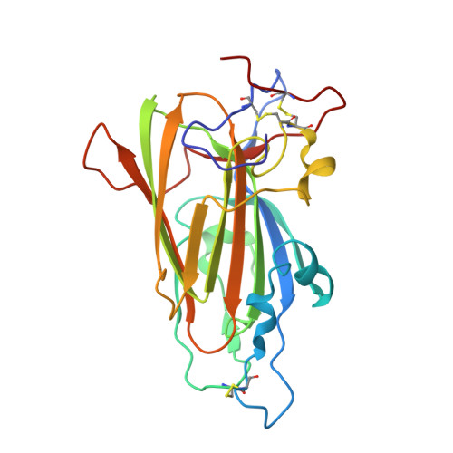
    4A3X

  • PubMed Abstract: 

    The yeast Candida glabrata represents the second major cause of clinical candidiasis cases in the world. The ability of this opportunistic pathogen to adhere to human epithelial and endothelial cells relies on the Epa adhesins, a large set of cell-wall proteins whose N-terminal domains are endowed with a calcium-dependent lectin activity. This feature allows the yeast cells to adhere to host cells by establishing multiple interactions with the glycans expressed on their cell membrane. The ligand-binding domain of the Epa1p adhesin, which is one of the best characterized in the Epa family, was expressed in Escherichia coli, purified and crystallized in complex with lactose. Sequence identity with the domain of another yeast adhesin, the Flo5p flocculin from Saccharomyces cerevisiae, was exploited for molecular replacement and the structure of the domain was solved at a resolution of 1.65 Å. The protein is a member of the PA14 superfamily. It has a β-sandwich core and a DcisD calcium-binding motif, which is also present in the binding site of Flo5p. However, Epa1p differs from this homologue by the lack of a Flo5-like subdomain and by a significantly decreased accessibility of the solvent to the binding site, in which a calcium ion still plays an active role in the interactions with carbohydrates. This structural insight, together with fluorescence-assay data, confirms and explains the higher specificity of Epa1p adhesin for glycan molecules compared with the S. cerevisiae flocculins.


  • Organizational Affiliation

    Research Group Structural Biology Brussels (SBB), Department of Bioengineering Sciences, Vrije Universiteit Brussel, Pleinlaan 2, 1050 Brussels, Belgium.


Macromolecules
Find similar proteins by:  (by identity cutoff)  |  3D Structure
Entity ID: 1
MoleculeChains Sequence LengthOrganismDetailsImage
EPA1P227Nakaseomyces glabratusMutation(s): 0 
UniProt
Find proteins for Q6VBJ0 (Candida glabrata)
Explore Q6VBJ0 
Go to UniProtKB:  Q6VBJ0
Entity Groups  
Sequence Clusters30% Identity50% Identity70% Identity90% Identity95% Identity100% Identity
UniProt GroupQ6VBJ0
Sequence Annotations
Expand
  • Reference Sequence
Oligosaccharides

Help

Entity ID: 2
MoleculeChains Length2D Diagram Glycosylation3D Interactions
beta-D-galactopyranose-(1-4)-beta-D-glucopyranose
B
2N/A
Glycosylation Resources
GlyTouCan:  G84224TW
GlyCosmos:  G84224TW
GlyGen:  G84224TW
Biologically Interesting Molecules (External Reference) 1 Unique
Experimental Data & Validation

Experimental Data

  • Method: X-RAY DIFFRACTION
  • Resolution: 1.65 Å
  • R-Value Free: 0.210 
  • R-Value Work: 0.172 
  • R-Value Observed: 0.174 
  • Space Group: C 2 2 21
Unit Cell:
Length ( Å )Angle ( ˚ )
a = 75.481α = 90
b = 105.453β = 90
c = 69.571γ = 90
Software Package:
Software NamePurpose
REFMACrefinement
XDSdata reduction
XDSdata scaling
BALBESphasing

Structure Validation

View Full Validation Report


Entry History 

Deposition Data

Revision History  (Full details and data files)

  • Version 1.0: 2012-02-22
    Type: Initial release
  • Version 1.1: 2012-03-07
    Changes: Other
  • Version 1.2: 2012-03-28
    Changes: Other
  • Version 2.0: 2020-07-29
    Type: Remediation
    Reason: Carbohydrate remediation
    Changes: Advisory, Atomic model, Data collection, Derived calculations, Non-polymer description, Other, Structure summary
  • Version 2.1: 2023-12-20
    Changes: Data collection, Database references, Refinement description, Structure summary