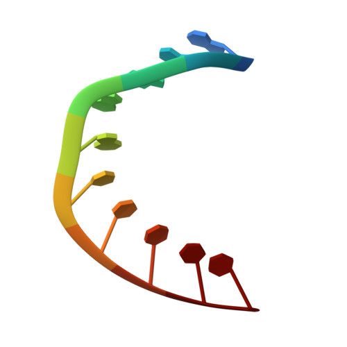
High-resolution A-DNA crystal structures of d(AGGGGCCCCT). An A-DNA model of poly(dG) x poly(dC).
Gao, Y.G., Robinson, H., Wang, A.H.(1999) Eur J Biochem 261: 413-420
- PubMed: 10215851
- Primary Citation of Related Structures:
440D, 441D - PubMed Abstract:
A-DNA conformation is favored by guanine-rich sequences, such as (dG)n x (dC)n, or under low-humidity conditions. Earlier A-DNA crystal structures revealed some conformational variations which may be the result of sequence-dependent effects and/or crystal packing forces. Here we report the high-resolution crystal structure of d(AGGGGCCCCT) in two crystal forms (either in the P212121 or the P6122 space group) to gain insights into the conformation and dynamics of the (dG)n x (dC)n sequence. The P212121 form has been analyzed using data to 1.1 A resolution by the anisotropic temperature factor refinement procedure of the SHELX97 program. Such analysis affords us with the detailed geometric, conformational and motional property of an A-DNA structure. The backbone torsional angles fall in a narrow range, except for the alpha/gamma angles which have two distinct combinations (gauche-/gauche+ or trans/trans). An A-DNA model of poly(dG) x poly(dC) has been constructed using the conformational parameters derived from the crystal structure of the P212121 form. In the crystal structure of the P6122 space group, the central eight base pairs of the decamer adopt A-DNA conformation with the two terminal nucleotides flipped out to form base pairs with the neighboring nucleotides. Comparison of the A-DNA structure of the same sequence from two different crystal forms, reinforced the conclusion that molecules crystallized in the same space group have a more similar conformation, whereas the same molecule crystallized in different space groups has different (local) conformations.
Organizational Affiliation:
Department of Cell & Structural Biology, University of Illinois at Urbana-Champaign, Urbana, IL 61801, USA.